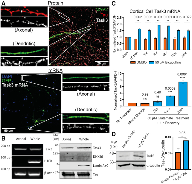
Figure 5.
Endogenous Task3 protein and mRNA are localised to distal neurites and upregulate in response to neuronal activity. (A, top) Immunocytochemistry of wild-type DIV10 embryonic murine cortical neurons stained for the endogenous dendritic and axonal markers, MAP2 (Green) and Tau (Red) respectively, as well as endogenous Task3 protein (white). (A, bottom) RNA Fluorescence in situ Hybridisation (FISH) of eGFP transfected DIV10 embryonic murine cortical neurons hybridised with anti-Task3 mouse probes showing endogenous Task3 mRNA from a singular GFP transfected neuron in distal neurites. (B) Western blot (left) and endpoint RT-PCR (right) analyses for specific protein and mRNA species’ from DIV10 primary embryonic cortical neurons cultured in Corning® Transwell® inserts for the isolation of whole neuron vs axonal proteins and mRNAs. (C) Total Task3 mRNA levels from DIV10 embryonic cortical neurons treated with 50 μM bicuculline or DMSO over a 24 h period (top), as well as with 50 μM glutamate for 30s, 1min, 2min or a no-treatment control media change with a 1h recovery period (bottom). (D) Western blotting of whole cortical cell lysates after 2 min treatment with 50 μM glutamate or no-treatment control media change with a 1 h recovery period (Left) with quantification of endogenous Task3 expression normalised to α-tubulin (Right). N = 4 biological replicates where error bars represent SEM. Statistical analyses were carried out by one-way ANOVA (C (bottom): (F(4, 10) = 90.60, P < 0.0001) with post-hoc Sidak's multiple comparison tests or by unpaired two-tailed t-tests; C (top) and D, P-values shown.

