Figure 1.
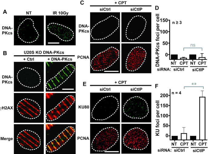
Ku and DNA-PKcs are differently removed from seDSBs. (A) Representative micrographs of DNA-PKcs foci detected by immunofluorescence in DNA-PKcs KO U2OS cells complemented with WT DNA-PKcs, untreated or irradiated with 10 Gy and incubated 5 min before being processed. (B) DNA-PKcs KO U2OS cells complemented or not with WT DNA-PKcs were microirradiaed with laser biphoton 5 min before being processed for DNA-PKcs and H2AX codetection by immunofluorescence. Representative micrographs are shown. (C, D) Representative micrographs (C) and quantification (D) of DNA-PKcs foci in replicating (PCNA positive) DNA-PKcs KO U2OS cells complemented with WT DNA-PKcs transfected by control or CtIP siRNA and treated for 1 h with DMSO or 1 μM CPT. (E, F) Representative micrographs (E) and quantification (F) of Ku foci in replicating U2OS transfected by control or CtIP siRNA and treated for 1 h with DMSO or 1 μM CPT. Error bars are s.d. NS, non-significant difference, as judged by t-test. White scale bars represent 10 μm.

