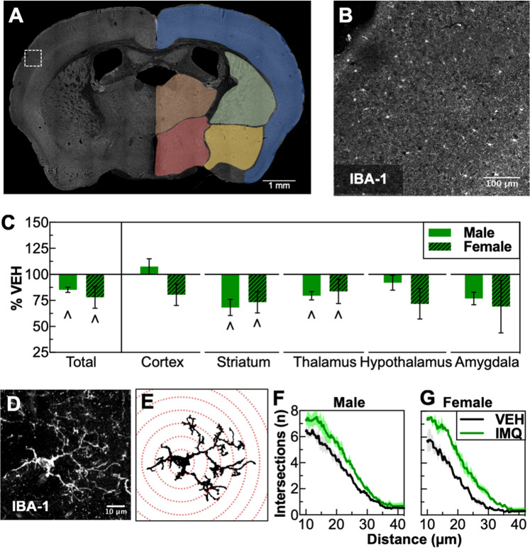
Fig. 4.
Prenatal TLR7 activation decreases microglia density and alters microglia morphology. a Example of a whole-section montage with different brain regions examined. b Inset of micrograph in a showing IBA-1 immunohistochemistry. c Decreased density of IBA-1-positive cells in several brain regions. d Maximum projection of a microglia cell from the dorsal striatum of a 7-µm z-stack from 19 images taken with a confocal microscope. e Graphical depiction of automated trace and Sholl analysis. f Results of Sholl analysis of microglia from dorsal striatum showing increased ramifications in males and (g) females. ∧P < 0.05 (main effect of VEH vs. IMQ), mean ± SEM

