Fig. 2.
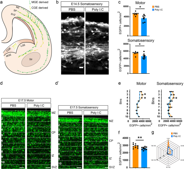
Early migratory deficits of GAD-EGFP+ neuroblasts due to maternal inflammation. a A schematic overview of the migratory routes taken by GAD+ neuroblasts during embryonic development [57, 58]. b, c Reduction in the density of EGFP+ neuroblasts migrating into the cortical intermediate zone at E14.5 as a result of maternal inflammation. Panel b shows somatosensory region of the developing cortex. Dotted box denotes the region of counting. Quantifications from the dotted boxes are shown in c (motor cortex: n = 8 each; somatosensory cortex: n = 8 each). d, e Superficial regions of the cortical column show a reduction in density of EGFP+ neuroblasts due to maternal inflammation. Cortical columns from developing motor (d) and somatosensory (d’) cortical regions were divided into ten equal-sized bins and the number of EGFP+ cells counted in each (with the exception of bin 1). Bin 10 represents the upper margin of the IZ. Densities in the graphs (e) show a reduction in bins 3–4. f Embryos exposed to maternal inflammation showed a decreased total density of migratory neuroblasts at E17.5. EGFP+ cells were counted in the cortical plate and intermediate zone across bins 2–10 (n = 10 each). g Radar plot showing that a greater percentage of neuroblasts in the cortical plate migrate laterally due to maternal inflammation as assessed by the angle of leading process. Angles were grouped into 30° bins and relative percentages were plotted. Each spoke represents 10%. Comparison of means by Student’s t-test in c, and f, t-test with Holm–Sidak correction for multiple comparison in e (mean ± SEM are shown, *p < 0.05, **p < 0.005). Scale bars: 50 µm

