Fig. 3.
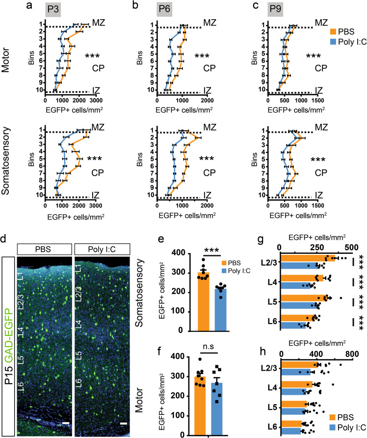
Maternal inflammation causes large-scale impairment in the distribution of GAD-EGFP+ neuroblasts and mature interneurons in the cortex. a–c Graphs showing a decrease in density of EGFP+ cells measured by counting EGFP+ cells in ten equally sized bins in the cortical plate of the presumptive motor and somatosensory cortices. Dotted lines represent the margins of IZ, CP, and MZ, respectively (P3 motor: F = 21.85, P3 somatosensory: F = 24.1, n = 8, 7 for PBS and poly I:C respectively; P6 motor: F = 21.72, P6 somatosensory: F = 19.59, n = 8, 8 for PBS and poly I:C respectively; P9 motor: F = 12.22, and P9 somatosensory: F = 13.77, n = 8, 10 for PBS and poly I:C respectively). d–h Significant reduction in the density of interneurons in the somatosensory but not in the motor cortex at P15. EGFP+ cells were quantified in the total cortical thickness as well as in each cortical layer. Panels in d show representative images of regions used for quantification. Graphs show the total (e, f) (n = 8 and 7 for PBS and poly I:C, respectively) as well as the layer-wise (g, h) reduction in EGFP+ cell density. Comparison of means by two-way ANOVA in a–c, Student’s t-test in e and f, and t-test with Holm–Sidak correction for multiple comparison in g and h) (mean ± SEM are shown, ***p < 0.0005). Scale bars: 50 µm (d)

