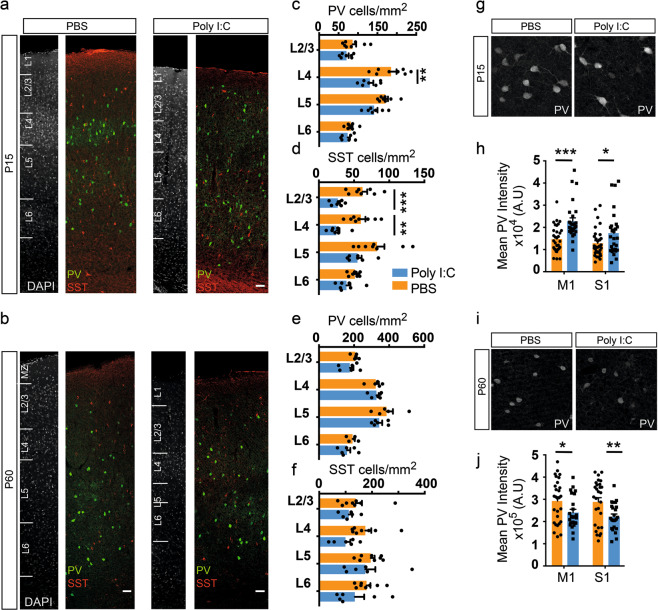
Fig. 4.
Interneuron subtype-specific effects of maternal inflammation. a–f Maternal inflammation resulted in a decreased density of PV and SST expressing interneurons at P15 but not at P60. Panels show representative images from P15 (a) and P60 (b). A significant decrease in PV+ interneurons could be seen in L4, while the difference was more pronounced in L2-4 for SST+ interneurons (c, d) (n = 8 and 7 for PBS and poly I:C, respectively). An appreciable but not statistically significant decrease could be seen at P60 (e, f). g–j Reciprocal changes in PV expression between P15 and P60 due to maternal inflammation. At P15, PV+ interneurons in both motor, and somatosensory cortical regions in maternal inflammation-exposed animals showed an increased expression of PV (h) (n = 8 and 7 for PBS and poly I:C, respectively) measured by the signal intensity in immunolabeled images (g). By P60 (i), this effect had reversed and instead a decreased expression was observed in both motor and somatosensory cortical regions (j) (n = 6 each). Statistical analysis by t-test with Holm–Sidak correction for multiple comparison in c–f, h, and j (mean ± SEM are shown, *p < 0.05, **p < 0.005, ***p < 0.0005). Scale bars: 50 µm (a, b)

