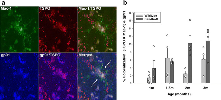
Fig. 6.
In situ colocalization of TSPO with gp91phox in murine brain microglia. a Representative triple labeled immunofluorescent confocal images in the thalamus of a 2-month old Sandhoff disease mouse at 60× with 1.6× zoom. Imaging confirmed that the TSPO signal colocalizes with the gp91phox signal in Mac-1 labeled microglia. b Quantification of percent colocalization of TSPO with gp91phox in microglia in the thalamus of Sandhoff disease mice compared to wildtype as a function of age. The data indicates that the TSPO-gp91phox interaction increases as a function of age and progression of neurological disease (age: F3,7 = 4.257, p = 0.017; genotype: F1,7 = 11.081, p = 0.003; age × genotype: F3,7 = 1.548, p = 0.232). Data are expressed as mean ± s.e.m. n = 4 independent animals and experiments per group, except for the 2-month old wildtype group which has n = 3 due to one animal being excluded as an outlier (> 3 standard deviations from the mean)

