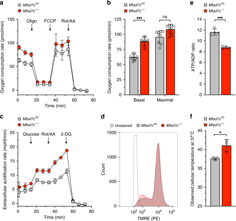
Fig. 3. Loss of Mfsd7c stimulates OCR, ECAR, and thermogenesis in BMDM.
Bone marrow-derived macrophages (BMDM) were derived from exon 2 floxed (Mfsd7cfl/fl) C57BL/6 mice and C57BL/6 mice with exon 2 deletion in macrophages (Mfsd7c−/−). a, b Representative OCR analysis from Mfsd7cfl/fl and Mfsd7c−/− macrophages as measured by Seahorse XF96e Analyzer and their responses to oligomycin (oligo), FCCP, and rotenone plus antimycin A (Rot/AA) (n = 3 technical replicates) (a), and their average basal and maximal OCR (b) (n = 9 independent experiments from 3 biological replicates). c Representative ECAR analysis of Mfsd7cfl/fl and Mfsd7c−/− macrophages and their responses to glucose, Rot/AA, and 2-deoxyglucose (2-DG) (n = 3 technical replicates). d Mitochondrial membrane potential of Mfsd7cfl/fl and Mfsd7c−/− macrophages analyzed with TMRE staining followed by flow cytometry (1 representative histogram picked from 3 biological replicates). e Cellular ATP/ADP ratio, (n = 4 biological replicates from average of 5 technical replicates). f Estimates of cellular temperature of Mfsd7cfl/fl and Mfsd7c−/− macrophages as measured by fluorescent thermoprobe (n = 3 biological replicates). p values were calculated using unpaired t-test (*p < 0.05, **p < 0.01, ***p < 0.005). Data are presented as mean value ± standard deviation.

