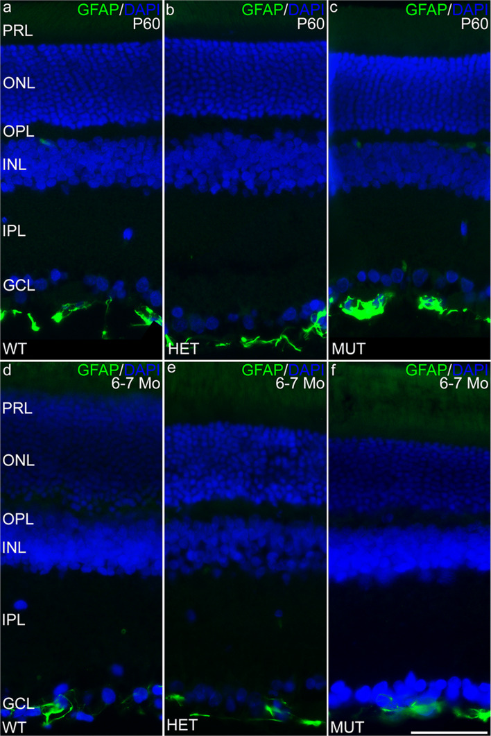
Fig. 8.
Müller cells in the SCA34-KI rat retina are not reactive. a–c Müller cells in the retina of P45 WT, HET, and MUT rats show little labeling for glial fibrillary acidic protein (GFAP) as is typical of Müller cells in the healthy retina. Labeling in blood vessels (bv) is non-specific. d–f Müller cells in the retina of 6–7-month-old (6–7 Mo) WT, HET, and MUT rats also show little labeling for GFAP, indicating the absence of glial reactivity associated with retinal degeneration. PRL, photoreceptor layer; ONL, outer nuclear layer; OPL, outer plexiform layer; INL, inner nuclear layer; IPL, inner plexiform layer; GCL, ganglion cell layer. Scale bars = 50 μm

