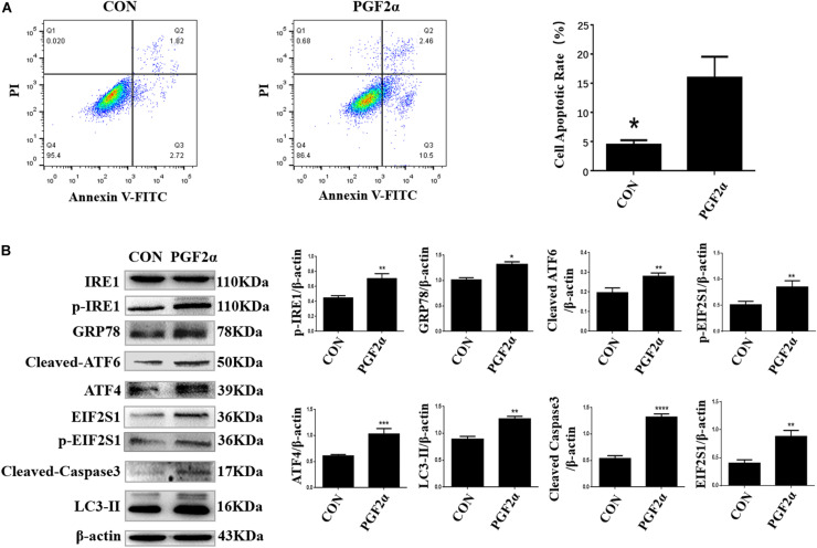
FIGURE 2.
PGF2α induces ER stress, autophagy, and apoptosis in goat luteal cells. (A) Cells cultured in medium with PGF2α (1μM) or isopycnic PBS for 24 h are stained with Annexin V-FITC/PI and analyzed by flow cytometry. B: Cells are treated with PBS or PGF2α for 24 h. The expression of IRE1, GRP78, ATF6, ATF4, and LC3 are detected by western blot analysis. The histogram shows relative protein expression displayed in (B) from three separate experiments. Data in the bar graph represent the means ± SEM of three independent experiments (N = 3). Means are compared with one-way ANOVA combined with Tukey’s post hoc tests. *p< 0.05, **p< 0.01, ***p< 0.001, ****p< 0.0001 compared to control.

