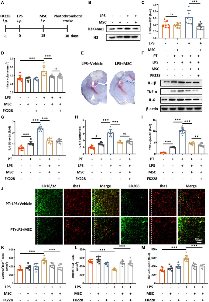
Figure 3.
Mesenchymal stem cells diminish LPS-induced innate immune memory and are protective in ischemic stroke. (A) Schematic diagram of experimental design. (B,C) Immunoblot (B) and quantitative analysis (C) showing that MSCs mitigate LPS-induced H3K4me1 elevation. Lipopolysaccharide induced significantly greater expression of H3K4me1, and this effect was mitigated by the administration of MSCs (n = 8 mice per group). (D) Quantitative analysis of infarct volume changes, showing that the LPS-induced augmentation of infarcts was diminished by MSCs (n = 8 mice per group). (E) Representative Nissl staining showing that LPS-induced elevation of infarct size was rectified by the MSC treatment. (F–I) Immunoblots (F) and quantitative analysis (G–I) showing that the LPS-induced increase in proinflammatory cytokines was rectified by MSCs. Interleukin 1β (G), IL-6 (H), and TNF-α (I) were significantly increased by LPS stimulation and diminished by MSCs. FK228 treatment further proved the specificity of MSCs in reacting this abnormal immune memory (n = 8 mice per group). (J–M) Representative immunofluorescence (J) and quantitative analysis (K–M) showing that the LPS-induced elevation of CD16/32-positive microglia (K) aggravated number of microglia surrounding parainfarct region (M), and down-regulation of CD206-positive microglia (L) was rectified by MSC treatment 7 days after photothrombotic stroke (n = 8 mice per group). Data were collected and analyzed with one-way ANOVA with Tukey multiple-comparisons test (ns, not significant; *p < 0.05; **p < 0.01; ***p < 0.001). Error bars, SD (“–” no corresponding treatment, “+” corresponding treatment, or administration).

