Figure 2.

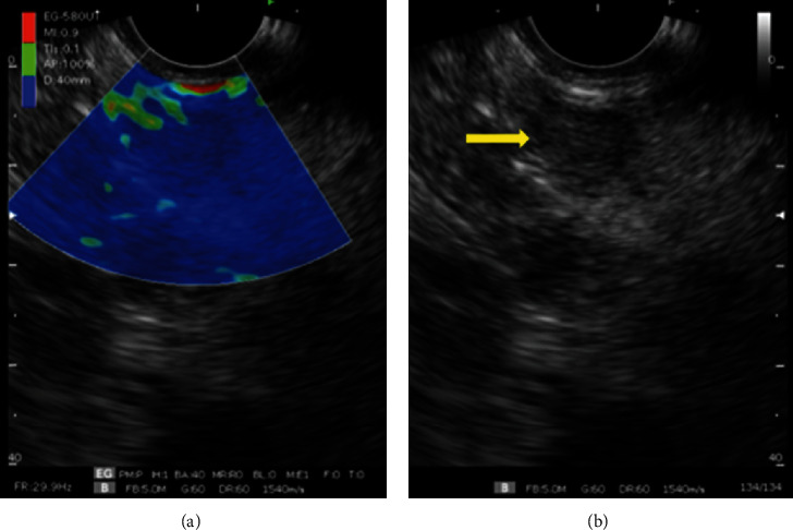
Elastography performed with a sectorial endoscopic ultrasound device. (a) Elastography of uncinate hardened process lesion (blue image). (b) Yellow arrow indicating hypoechoic lesion of uncinate process.

Elastography performed with a sectorial endoscopic ultrasound device. (a) Elastography of uncinate hardened process lesion (blue image). (b) Yellow arrow indicating hypoechoic lesion of uncinate process.