Figure 3.
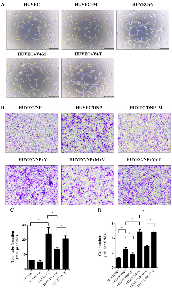
Tube formation and cell migration of HUVECs. HUVECs were pretreated with melatonin (10 µM) or TGF-β1 (10 ng/ml) for 4 h and then incubated with NP-conditioned medium and 25 ng/ml VEGF for 6 h. (A) Tube formation was observed under a light microscope. Scale bar, 50 µm. (B) HUVECs were co-cultured with NP or DNP and incubated with melatonin (10 µM) or TGF-β1 (10 ng/ml) for 24 h. After fixation and staining, HUVEC migration was observed under a light microscope. Scale bar, 50 µm). (C) Tube formation and (D) cell migration of HUVECs was quantified. Values are expressed as the mean ± standard deviation. *P<0.05. DNP, degenerative nucleus pulposus cells; HUVECs, human umbilical vein endothelial cells; M, melatonin; NP, nucleus pulposus cells; T/TGF-β1, transforming growth factor-β1; V/VEGF, vascular endothelial growth factor.

