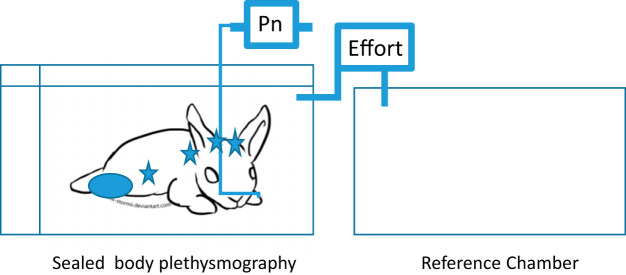
Fig. 2.
The experimental setup. The drawing shows a schema of the experimental setup. The drawing illustrates the body box setup with the recording and reference chamber. The stars represent the general electrode placement for EEG, oximeter, and ECG. Pressure at the nares (Pn) by nasal cannula was to monitor flow. Effort was recorded as the pressure of the box is referenced to an empty box of the same size.

