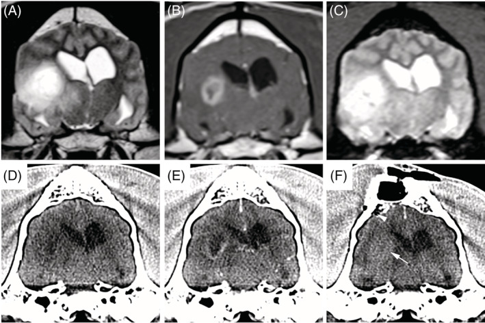
FIGURE 2.

Pneumocephalus and iatrogenic intratumoral hemorrhage after stereotactic brain biopsy in a case dog that developed adverse events. Transverse prebiopsy MRI (A, precontrast T2W; B, postcontrast T1W; and C, T2*gradient echo) and transverse stereotactic planning CT (D, precontrast; E, postcontrast) of a high‐grade astrocytoma in the temporal lobe demonstrating T2W heterogeneous signal (A) and marked ring enhancement (B). There is no evidence of intracranial hemorrhage on the prebiopsy images (A‐E), while hyperattenuating intratumoral hemorrhage (F; arrow) and a small focus of subarachnoid gas (arrowhead) are visible on the postbiopsy CT scan
