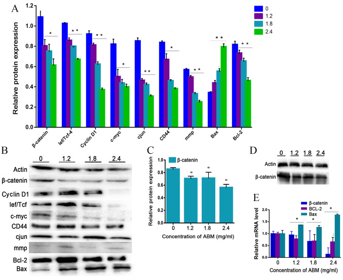
Figure 4.
AbM induces apoptosis of K562 cells. K562 cells were treated with different concentrations of AbM (0, 1.2, 1.8 and 2.4 mg/ml) for 48 h and the expression of apoptosis-associated genes and proteins was determined using western blot analysis for (A and B) 24 h (*P<0.05 and **P<0.001) or (C and D) 48 h and (E) reverse transcription-quantitative PCR. The relative expression levels of β catenin, Bax and Bcl-2 genes in K562 cells were quantified according to the gene expression levels of actin. The relative expression levels of cyclin D1, Lef/Tcf, c-myc, β catenin, CD44, c-Jun, mmp, Bax and Bcl-2 proteins in K562 cells were quantified according to the protein expression levels of actin. Data are representative of three independent experiments and are expressed as the mean ± SD. *P<0.05 vs. 0 mg/ml.

