Figure 1.
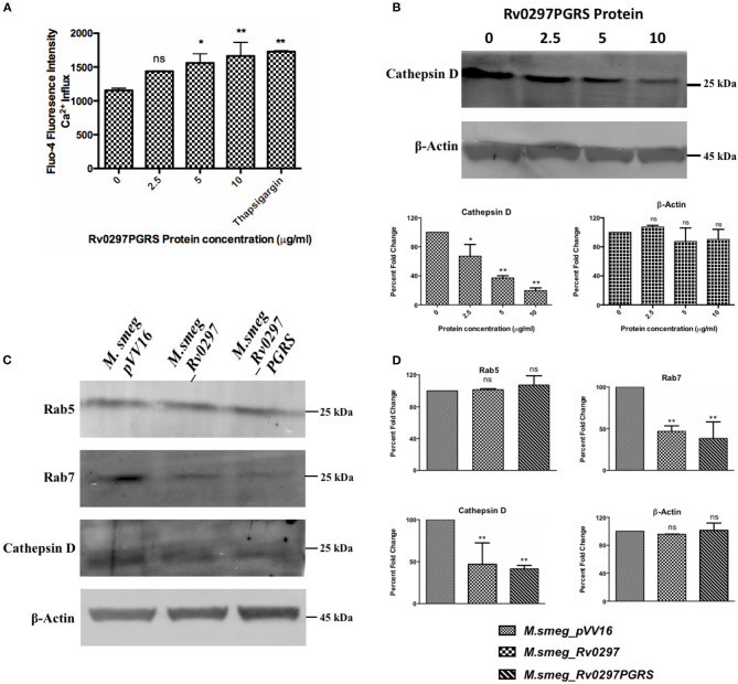
Rv0297 downregulated the phagolysosomal acidification. Ca2+ release from Rv0297PGRS-stimulated (for 30 h) THP-1 cells measured by Fluo-4 dye (A). Thapsigargin (1 mM) was used as the positive control. (B,C) Western blots depicting the downregulation of the early and late phagosomal markers (Rab5, Rab7, and cathepsin D). (B) The levels of cathepsin D were assessed upon stimulation of the macrophages with Rv0297PGRS for 30 h. (C) Levels of the early and late phagosomal markers were assessed in THP-1 macrophages infected with M.smeg_VC, M.smeg_Rv0297, and M.smeg_Rv0297PGRS for 48 h. To ensure the equal loading of lysates, β-actin levels were loaded and immunoblotted. The data shown are representative of three independent experiments. (D) Densitometric analysis of the Western blots depicted in (C). *P < 0.05, **P < 0.01, and P > 0.05 (ns).

