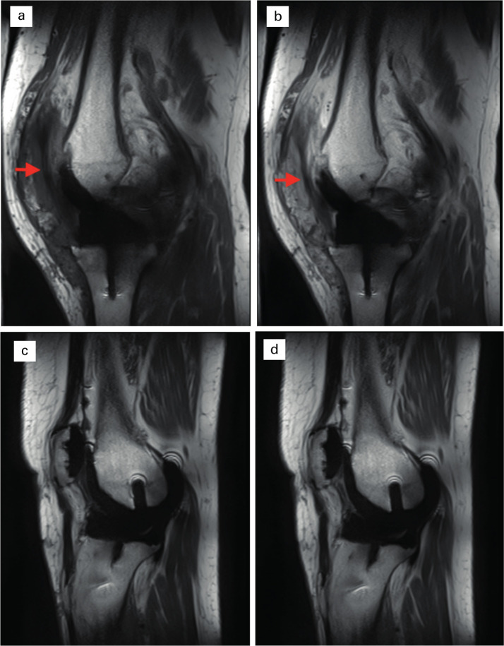
Fig. 3.
Fibrotic tissue in the infrapatella region. Sagittal pre- and post-contrast T1 images comparing fibrotic patient (a) and (b) with non-fibrotic (c and d). Fibrotic tissue (arrows) is identified in the infrapatella region in a and b, extending underneath the patella between the infra- and suprapatella pouches. No such band of tissue is seen in a healthy TKA (c and d).

