FIGURE 4.

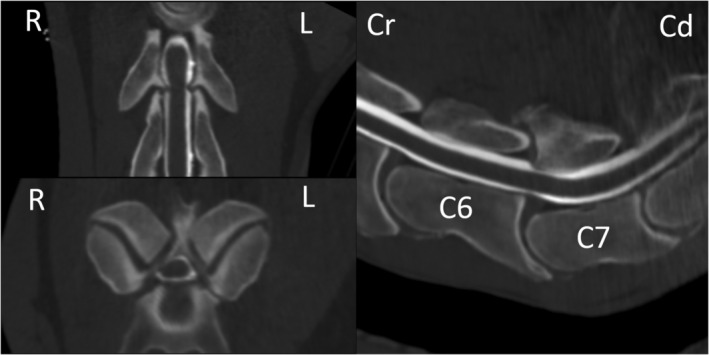
Computed tomographic (CT) myelographic 3D multiplanar reconstruction at the level of C6‐C7 in case 33. Note the profound narrowing of the contrast media column surrounding the spinal cord at this level

Computed tomographic (CT) myelographic 3D multiplanar reconstruction at the level of C6‐C7 in case 33. Note the profound narrowing of the contrast media column surrounding the spinal cord at this level