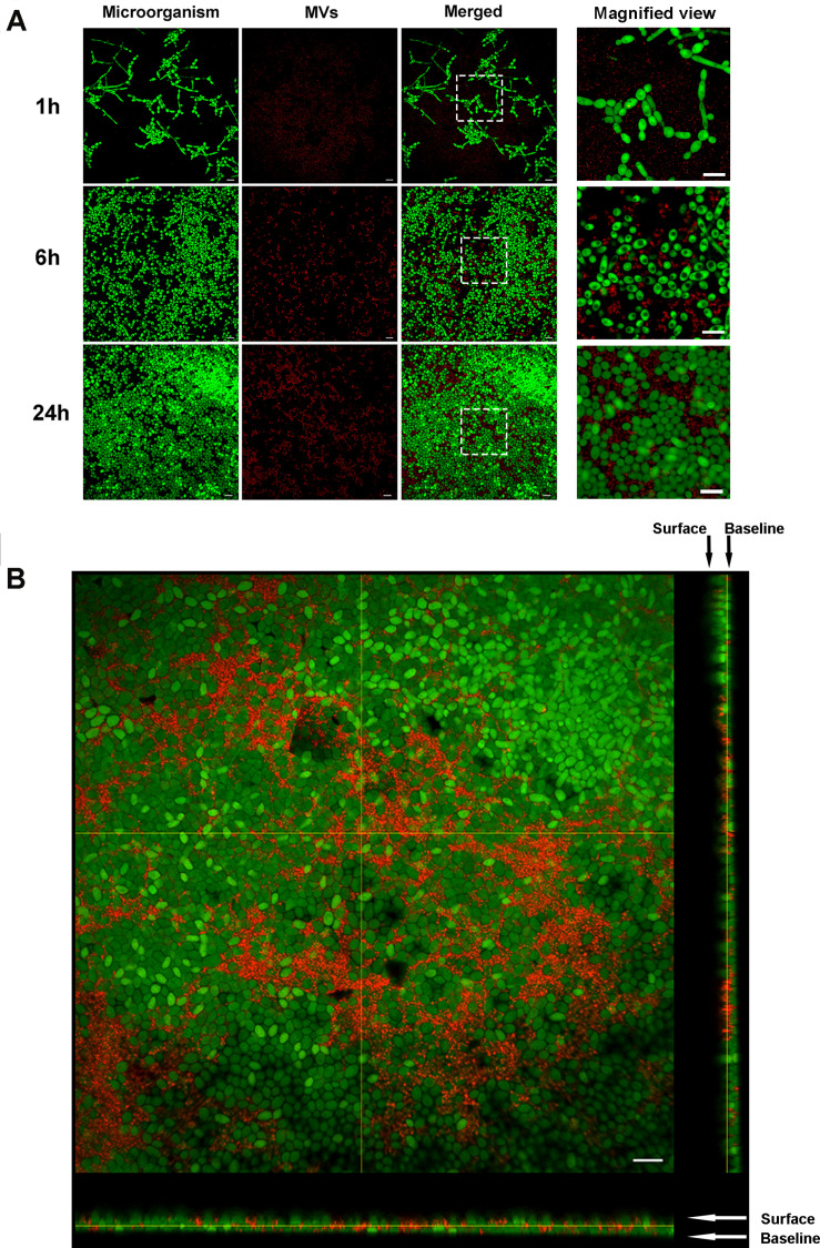
FIGURE 4.
Fluorescent labeling of MVs and tracing. PKH26 (in red)-stained MVs were incubated with C. albicans at different time points. The C. albicans cells were stained with SYTO-9 (in green). (A) Representative CLSM images of C. albicans biofilms at 1, 6, and 24 h, the white boxes indicate the magnified viewing area. (B) Three-dimensional reconstructions of C. albicans 24-h-old biofilms. Magnification, 60×; scale bar, 20 μm. CLSM images showed that S. mutans MVs were located in the C. albicans biofilm extracellular matrix.

