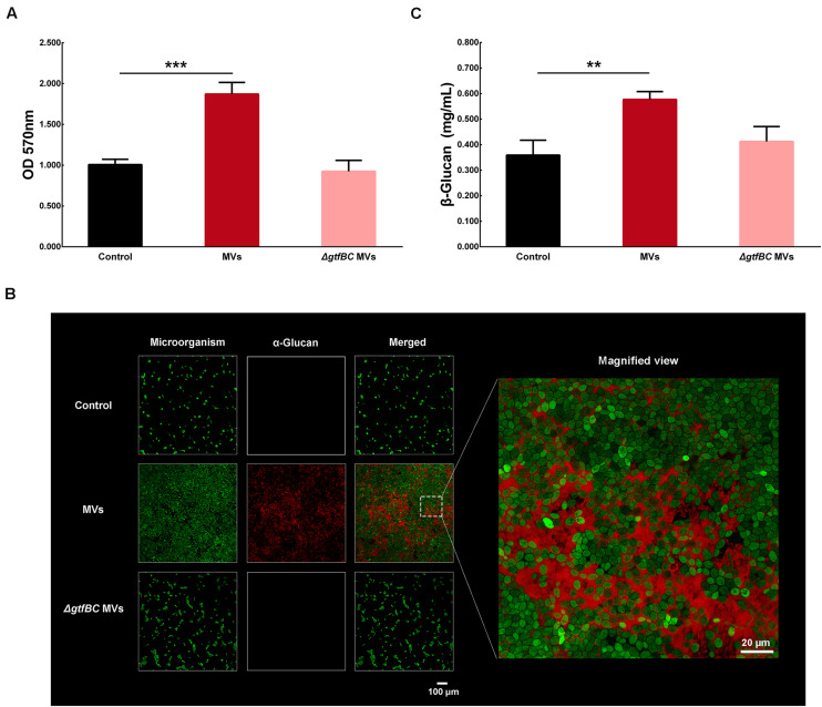
FIGURE 6.
C. albicans biofilm formation by S. mutans MVs. (A) Crystal violet assay of C. albicans 24-h biofilm formation in conditions with S. mutans MVs and S. mutansΔgtfBC mutant MVs. (B) CLSM images taken at 20 × magnification. α-Glucans were detected with an Alexa Fluor 647 dextran conjugate (in red), and the C. albicans cells were stained with SYTO-9 (in green). The white box indicated the magnified view area. (C) β-glucans concentration of C. albicans biofilm. The experiments were performed in three distinct replicates, and the data are presented as the means ± SD, ***P < 0.001, **P < 0.01 vs control group.

