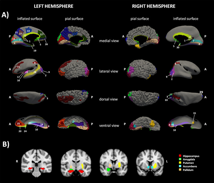
Figure 1.
Sub-network cortical/subcortical labels. The figure shows the sub-network of significant difference between the two groups of children obtained with network-based statistic (NBS). (A) shows the cortical nodes while (B) shows basal ganglia belonging to the sub-network. 1=L and R vPCC G, 2=L and R Middle occipital G, 3=L and R Superior occipital G, 4=L and R Lingual part of the medial occipito-temporal G, 5=L and R Subcallosal G, 6=L and R Occipital Pole, 7=L and R Calcarine S, 8= L and R Intraparietal and tansverse parietal S, 9=L and R Middle Occipital and Lunatus S, 10=L and R Collateral and Lingual S, 11=L and R Pericallosal S, 12=L ACC G and S, 13=L pMCC G and S. 14=L Cuneus G, 15=L Middle Frontal G, 16= L Long Insular G and central S of the Insula, 17=L Short Insular G, 18=L Fusiform G, 19=L Parahippocampal part of the medial occipito-temporal G, 20=L Orbital G, 21=Precuneus G, 22= L Inferior Segment of the Circular S of the Insula, 23=L Anterior Transverse Collateral S, 24=L Medial Orbital (Olfactory) S, 25= L Orbital (H Shaped) S, 26=L Suborbital S, 27=L Subparietal S, 28=L Superior Temporal S, 29=R Transverse Frontopolar G and S, 30=R Planum polare of the Superior Temporal G, 31=R Temporal Pole, 32=R Posterior Transverse Collateral S, 33= R Superior occipital and Transverse Occipital S. G=Gyrus/i, S=Sulcus/i; R=right hemisphere, L=left hemisphere, ACC=anterior cingulate cortex, pMCC=middle posterior cingulate cortex, vPCC=ventral part of the posterior cingulate cortex.

