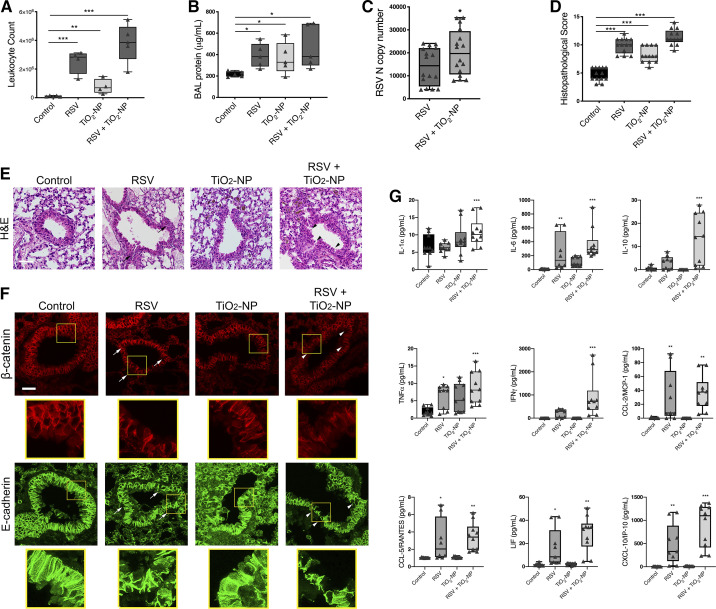
Fig. 8.
Exposure to titanium dioxide nanoparticles (TiO2-NP) enhances respiratory syncytial virus (RSV)-induced barrier disruption in vivo. Mice were intranasally inoculated with TiO2-NP (2 mg/kg), followed immediately by RSV. On day 4, lungs and bronchoalveolar lavage (BAL) were harvested and analyzed for white blood cell infiltration (A), protein (B), RSV nucleocapsid (N) copy number in lung homogenates (C), and histopathological score and photomicrographs of hematoxylin-eosin (H&E)-stained lung tissue section (D and E), and disruption of apical junctional complex (AJC) proteins β-catenin and E-cadherin (F). Arrows point to areas of peribronchial inflammation (black) and AJC disruption (white) after exposure to RSV or TiO2-NP; arrowheads point to areas of exaggerated inflammation (black) and AJC disruption (white) after coexposure. Scale bar = 40 μm. Data are presented as means ± SE; n = 10 mice per group, *P < 0.05, **P < 0.01, and ***P < 0.001 vs. control or TiO2-NP groups. Exposure to TiO2-NP modifies proinflammatory cytokine expression during RSV infection in vivo (G). Cytokine concentrations of BAL of mice exposed to rrRSV, TiO2-NP, or a combination were determined. Interleukin (IL)-1α, IL-6, IL-10, tumor necrosis factor-α (TNFα), interferon-γ (IFNγ), monocyte chemoattractant protein (MCP)-1, RANTES, LIF, and IFNγ-induced protein 10 (IP-10) protein concentrations were higher in BAL fluid of mice exposed to both TiO2-NP and RSV. Data are presented as means ± SE; n = 5 mice per group, of 2 independent experiments, *P < 0.05, **P < 0.01, ***P < 0.001 vs. control group.

