Figure 5.
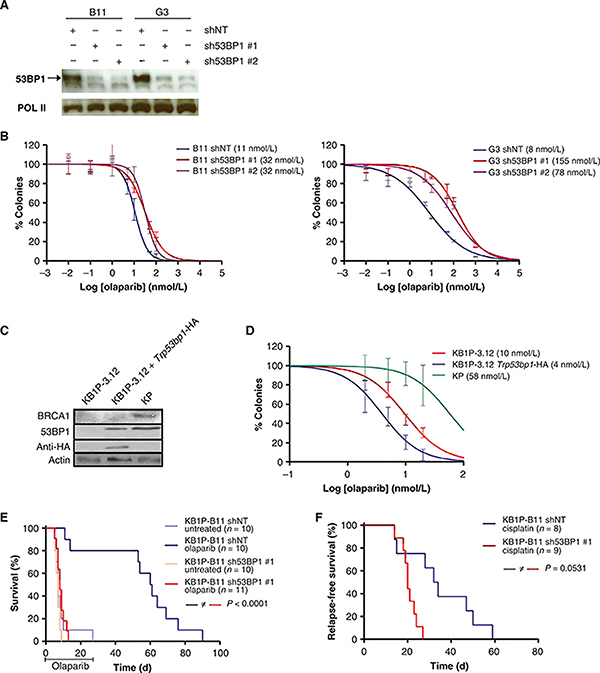
The effect of 53BP1 loss on olaparib sensitivity. A, Western blot analysis showing 53BP1 levels in KB1P-B11 and KB1P-G3 cells that express a nontargeting hairpin (NT) or a hairpin against Trp53bp1. B, clonogenic assay with olaparib. The IC50 is indicated between brackets. C, Western blot analysis showing the reconstitution of 53BP1 in 53BP1-deficient KB1P-3.12 cells. KP cells are used as positive control for the BRCA1 Western blot analysis. D, clonogenic assay of 53BP1-negative KB1P-3.12 cells, h53BP1-reconstituted KB1P-3.12 cells, and BRCA-proficient KP cells. E, overall survival of mice with a 53BP1-positive (shNT) or -negative (sh53BP1) tumor treated with one regimen of olaparib daily for 28 days and the untreated control mice. 53BP1 expression in these tumors is shown in Supplementary Fig. S7C. F, relapse-free survival of mice with a 53BP1-positive (shNT) or negative (sh53BP1) tumor treated with one dose of cisplatin. The Gehan–Breslow–Wilcoxon P values are indicated.

