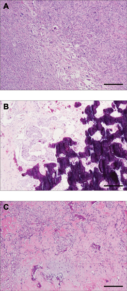
Figure 1 -. Histologic characteristics of metaplastic breast carcinomas (MBCs) with osseous differentiation.

Representative photomicrographs of hematoxylin and eosin (H&E)-stained MBCs included in this study. (A) MTC26 displaying areas of spindle cell morphology with cells arranged in a storiform pattern. (B) MTC27 also displays the intervening epithelial differentiation around osseous areas. (C) MTC28 classified showing osseous and chondroid differentiation in a myxoid background. Scale bar, 500 μm (A, C), 200 μm (B).
