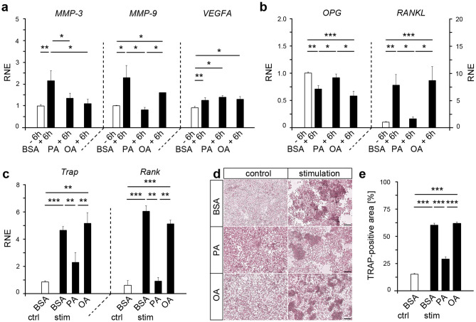
Figure 3.
Fatty acid cultivation of mechanically stressed HPdLF affect genes relevant for extracellular matrix degradation as well as influence osteoclast differentiation. (a) Quantitative expression analysis of extracellular matrix (ECM)-remodeling genes MMP-3 and MMP-9 as well as VEGFA coding for an vascularization factor in HPdLF treated with palmitic acid (PA) or oleic acid (OA) and stimulated with compressive force over six hours compared to unstimulated BSA controls. (b) Quantitative expression analysis of osteoclastogenesis-relevant genes OPG and RANKL in HPdLF treated with fatty acids and stimulated with compressive force over six hours compared to unstimulated BSA controls. (c) Quantitative expression analysis of osteoclast-related genes Trap and Rank in mouse osteoclasts stimulated with the media supernatant of mechanically compressed HPdLF cultured in fatty acids compared to BSA (stim BSA, stim PA, stim OA) and only BSA-containing media as control (ctrl BSA). The results are normalized to ctrl BSA. (d) Representative microphotographs of TRAP-staining of osteoclasts stimulated with the medium supernatant of fatty acid treated and mechanically compressed HPdLF quantified as TRAP-positive area of the total area in (e). *P < 0.05; **P < 0.01; ***P < 0.001; One-Way ANOVA and post-hoc test (Tukey). Photographs were analyzed with Fiji software (https://imagej.net/Fiji). Scale bars: 50 μm in (c) Ctrl, control; BSA, bovine serum albumin; HPdLF, human periodontal ligament fibroblast; OA, oleic acid; PA, palmitic acid; RNE, relative normalized expression; stim, stimulation.

