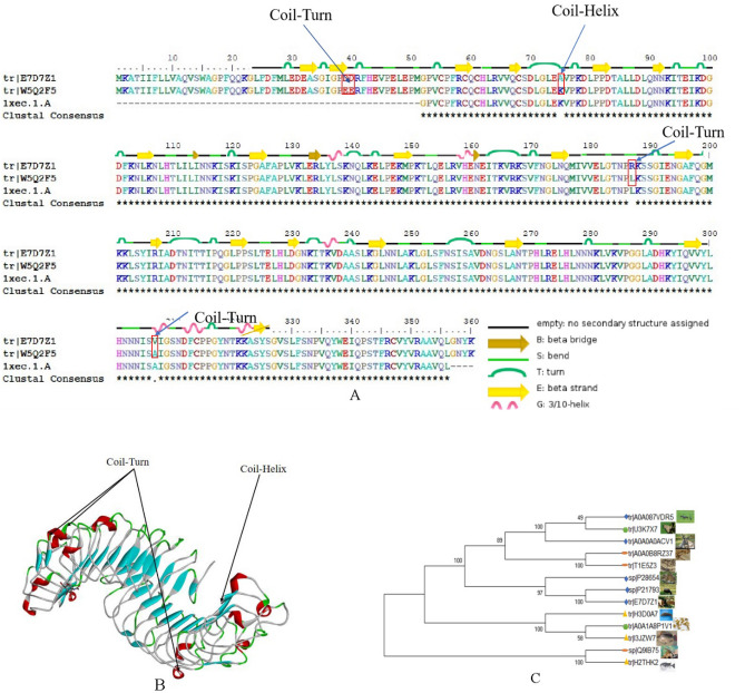
Figure 5.
Protein Sequence alignment, 3D modeling and phylogenetic analysis of Decorin from SDAPR. (A) A presumed sequence W5Q2F5 aligned with the model Decorin. At the bottom of columns, asterisks (*) show conserved positions, colons (:) show conserved substitutions and points (.) show non-conserved substitutions. Grey line, green bend, blue banded arrowhead and red solenoid represent coil, turn, sheet and helix, respectively. Different fragments are framed by red lines. (B) 3D modeling was simulated using the template Decorin by SWISS-MODEL and viewed by Discovery Studio 4.5. The colors grey, green, blue and red represent coils, turns, sheets and helices, respectively. Different fragments are indicated by red arrows. (C) Phylogenetic tree constructed using putative Decorin and 13 other sequences from different species using MEGA 7 with the Neighbor-Joining method.

