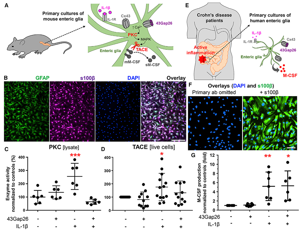
Figure 6. Glial M-CSF Production Is Regulated by Cx43, PKC, and TACE.

Data from in vitro experiments with a primary mouse (A–D) and human (E–G) enteric glia.
(A) Primary cultures of mouse enteric glia were derived from the colon myenteric plexus. Schematic showing proposed mechanisms underlying IL-1β-induced M-CSF release. IL-1R, IL-1 receptor; Cx43, Cx43 hemichannel; 43Gap26, Cx43 mimetic peptide that binds to extracellular loops of Cx43 and blocks Cx43 hemichannels; ↑Ca2+, increase in cytosolic calcium; PKC, protein kinase C; MAPK, a mitogen-activated protein kinase; TACE, tumor necrosis factor α-converting enzyme; mM-CSF and sM-CSF, membrane-bound and soluble M-CSF.
(B) Representative images of mouse enteric glia cultures labeled with markers of enteric glia (GFAP, green, and s100β, magenta) and counterstained with the nuclear marker DAPI (blue). Scale bar, 100 μm.
(C and D) Quantification of PKC and TACE activity in primary cultures of mouse enteric glia stimulated overnight with IL-1β (+ IL-1β, 1 ng/mL) in the presence or absence of 43Gap26 ±43Gap26, 100 μM). (C) PKC activity in cell lysates was normalized to total protein concentration in the cell lysate and to mean of untreated controls. (D) TACE activity in live-cell suspensions was normalized to cell density and to untreated controls that originated from the same colon. *p = 0.0342; ***p = 0.0008; ANOVA followed by Dunnett’s multiple comparisons test. Data are shown as mean ± SD, n = 6 and 11–13 mice, respectively.
(E) Human enteric glial cells were cultured from segments of the intestine harvested from individuals undergoing resections for Crohn disease. Cultured gliawere incubated with IL-1β to stimulate the production of proinflammatory cytokines, and a subset of cultures was co-incubated with 43Gap26.
(F) Representative images of human enteric glia derived from the distal colon. Images show labeling for s100β (glia, green) and DAPI (nuclei, blue) in samples where the primary anti-s100β antibody was omitted (left) or fully stained (right). Scale bar, 100 μm.
(G) Quantification of M-CSF in supernatants from human enteric glial cultures after IL-1β stimulation (+ IL-1β, 1 ng/mL) and co-incubation with 43Gap26 (+ 43Gap26, 100 μM). Raw M-CSF concentrations were normalized to cell density and to untreated controls that originated from the same culture. *p = 0.0103; **p = 0.0097; ANOVA followed by Dunnett’s multiple comparisons test. Data are shown as mean ± SD, n = 3–4 patients (duplicate cultures derived from involved or noninvolved regions). Besides, proinflammatory signals change the expression of connexin genes in mouse and human enteric glia (Figure S6).
