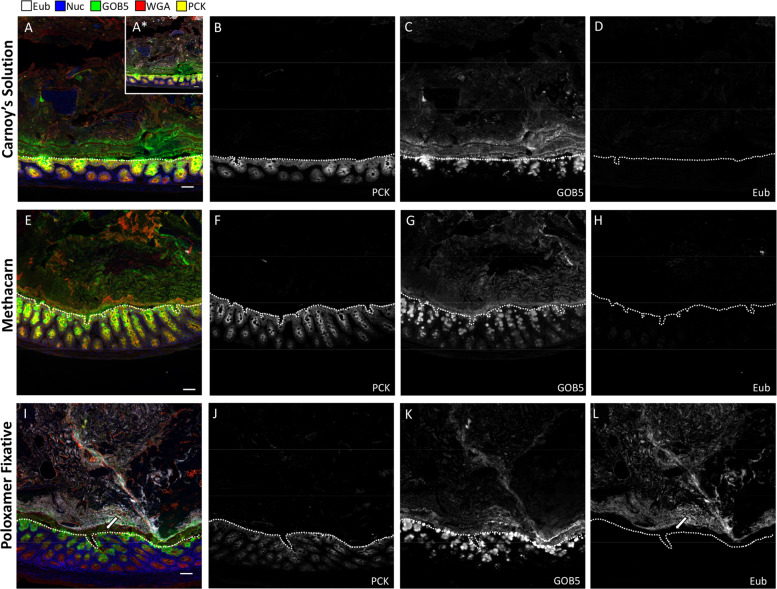
Fig. 3. Poloxamer fixation enables concurrent analysis of microbes and tissues, as compared with Carnoy’s and Metharn fixation.
IF of proteins coupled to bacterial FISH over multiple rounds of MxIF, with universal bacterial probe (Eub, white), nuclei (blue), GOB5 (green), lectin (WGA, red), and pan-cytokeratin (PCK, yellow). Selected individual channels (PCK, GOB5, Eub) to visualize epithelium, mucus, and bacteria, with the EUB probe having the same exposure times and gains to compare signal-to-noise in the two conditions. a–d Carnoy’s solution, e–h Methacarn, and i–l Poloxamer fixative were used for processing the same distal colonic tissue. A* ×10 gain was used to detect Eub probe in the Carnoy’s condition. White dotted lines represent epithelial borders. Scale bars = 50 µm.

