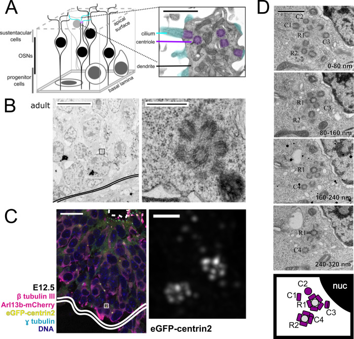
Fig 1. Location of rosette structures and free centrioles in adult and embryonic mouse olfactory epithelium.
(A) Schematic of the olfactory epithelium. The inset shows an OSN dendrite from adult mouse imaged by TEM with pseudocolored cilia and centrioles. Inset scale bar = 1 μm. (B) TEM image of wild-type adult mouse olfactory epithelium. Double solid line marks the basal lamina. Box shows the approximate location of the inset in the panel to the right. The panel on the right shows an inset of a centriole rosette in cross section, near the basal lamina. Scale bar = 10 μm. Inset scale bar = 0.5 μm. (C) Fluorescence image of embryonic olfactory epithelium at E12.5 in mice expressing eGFP-centrin2 to mark centrioles, as well as Arl13b-mCherry to mark cilia. The maximum projection inset shows a deconvolved image of 2 rosette-like centriole clusters from a cell positive for β tubulin III, near the basal lamina. Dashed line marks the apical surface of the olfactory epithelium. Double solid line marks the basal lamina. Box shows the location and orientation of the inset. Scale bar = 20 μm. Inset scale bar = 1 μm. (D) TEM images of wild-type adult mouse olfactory epithelium in serial sections. The images show 2 centriole rosettes, R1 and R2, and free centrioles, C1-4. Section sequence is indicated in the bottom right of each panel. The bottom panel summarizes the locations of centrioles in all 4 panels, where new centrioles are shown in purple and mother centrioles are shown in gray. Scale bar = 1 μm. See S1 Fig for additional details. Arl13b-mCherry, ADP-ribosylation-factor-like GTPase 13b conjugated to mCherry; eGFP, enhanced green fluorescent protein; nuc, nucleus; OSN, olfactory sensory neuron; TEM, transmission electron microscopy.

