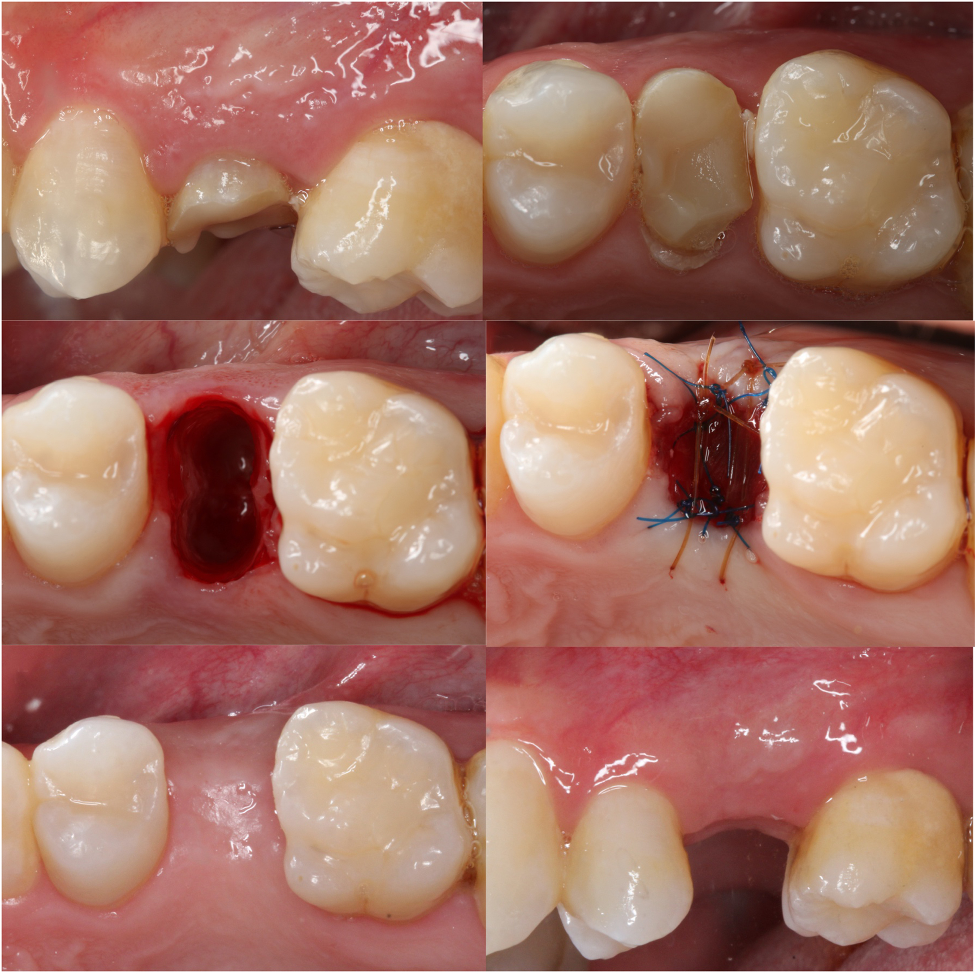
Figure 2.

Clinical images of the procedures performed for the collagen matrix group. A. Buccal view prior to the extraction of tooth #13; B. Occlusal view; C. Extraction socket after minimally invasive extraction; D. Extraction socket grafted with deproteinized bovine bone and covered with collagen matrix; E. Occlusal view 6 months post-extraction; F. Buccal view 6 months post-extraction.
