Figure 1.
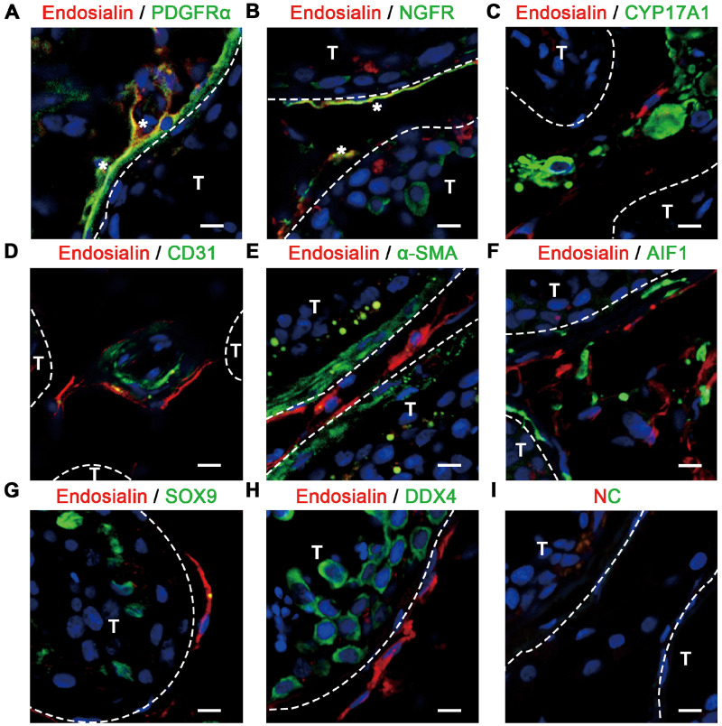
Expression pattern of endosialin in human testes, as shown by immunofluorescence. (A–C) Endosialin was co-expressed with the reported SLCs markers PDGFRα (A) and NGFR (B) but did not co-localize with the LCs marker CYP17A1 (C) in the interstitial region. Co-expression is marked with white asterisks in (A) and (B). (D, E) Endosialin+ cells were located in the perivascular region (endothelial cells are marked with CD31) (D) and peritubular region (peritubular myoid cells are marked with α-SMA) (E) of the human testes. (F–H) Endosialin-expressing cells did not express the macrophage marker AIF1 (F), the Sertoli cell marker SOX9 (G) or the germ cell marker DDX4 (H). (I) The negative control stained in the absence of the primary antibodies is shown. Nuclei were counterstained with DAPI. T and interstitium were indicated by dotted lines. The scale bars denote 10 μm. The images are representative of results obtained from patient samples (n = 3). AIF1, allograft inflammatory factor 1; CYP17A1, cytochrome P450 family 17 subfamily A member 1; DAPI, 4,6-diamidino-2-phenylindole; DDX4, DEAD-box helicase 4; LCs, Leydig cells; NC, negative control; NGFR, nerve growth factor receptor; PDGFRα, platelet-derived growth factor receptor α; SLCs, stem Leydig cells; SOX9, SRY-box transcription factor 9; T, seminiferous tubules; α-SMA, alpha-smooth muscle actin.

