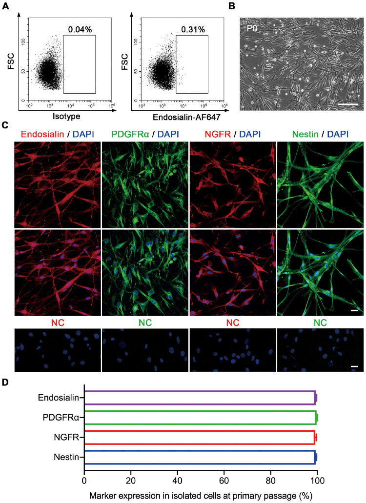
Figure 2.
Isolation and identification of endosialin+ cells from human testes. (A) Endosialin+ cells were isolated by FACS from adult human testes (n = 4). Left: isotype controls. Right: stained samples. (B) Representative phase-contrast micrograph of endosialin+ cells at P0 (n = 4). The scale bar denotes 200 μm. (C) Endosialin+ cells expressed the SLCs markers PDGFRα, NGFR and Nestin (n = 3). The nuclei were counterstained with DAPI. NC stained in the absence of the primary antibody. The scale bars denote 25 μm. (D) The percentages of endosialin+, PDGFRα+, NGFR+ and Nestin+ cells among isolated cells were analysed. The data are presented as the mean ± SEM (n = 3). FACS, fluorescence-activated cell sorting; FSC, forward scatter; P0, primary culture.

