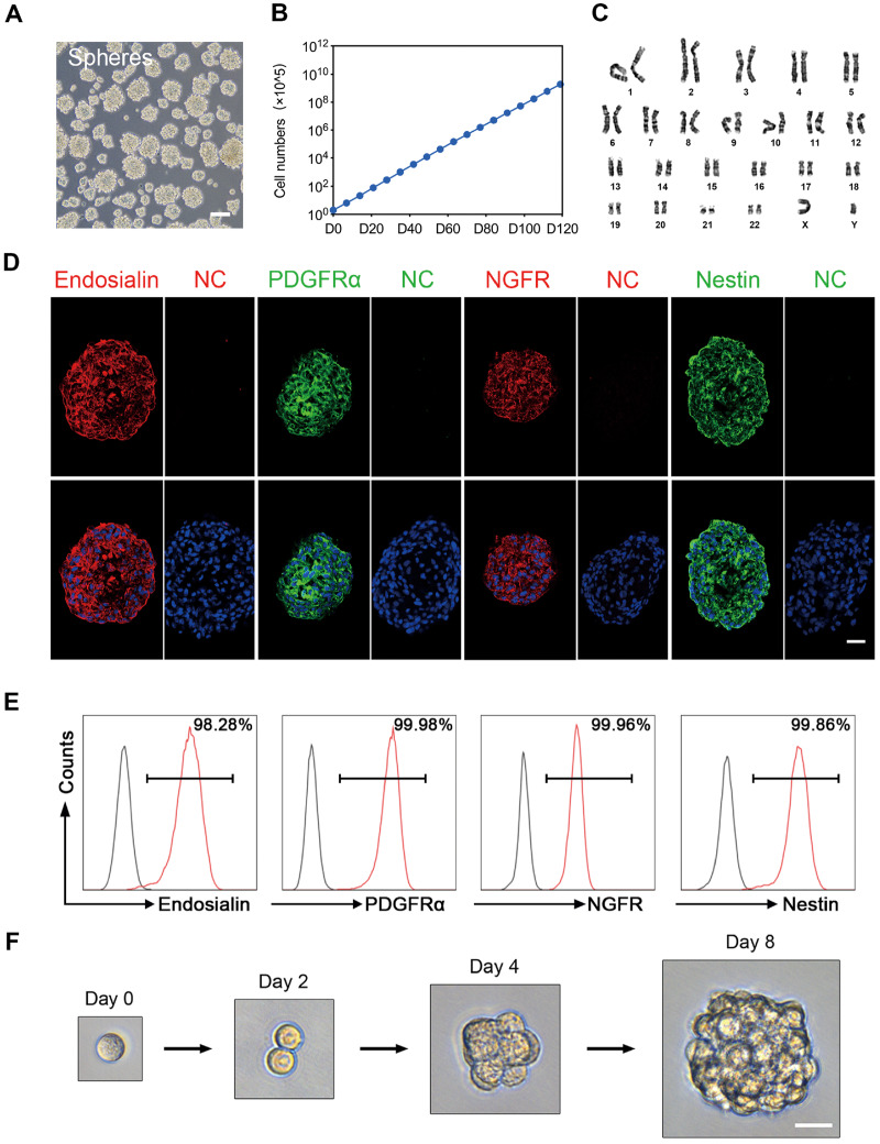
Figure 3.
Proliferation and self-renewal capacity of endosialin+ cells. (A) Phase-contrast micrograph of floating spheres at P3 growing from human testicular endosialin+ cells in serum-free expansion medium (n = 3). The scale bar denotes 100 μm. (B) Proliferation curve of endosialin+ cells cultured in expansion medium over different passages. The initial cell count was 2 × 10⁵. The data are expressed as the mean ± SEM (n = 3). (C) Karyotypic stability of expanded endosialin+ cells up to P20 was analysed. (D) Immunofluorescence staining for the indicated markers (Endosialin, PDGFRα, NGFR and Nestin) in endosialin+ cell spheres. The nuclei were counterstained with DAPI. NC stained in the absence of the primary antibody. The scale bars denote 25 μm. (E) FACS analysis for detection of the indicated markers (Endosialin, PDGFRα, NGFR and Nestin) in endosialin+ cell spheres (n = 3). (F) Representative images of a clonal sphere growing from a single cell, as observed using bright-field microscopy (n = 3). The scale bar denotes 25 μm. P3, passage 3; P20, passage 20.

