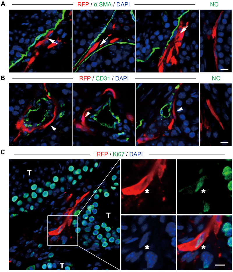
Figure 5.
Localization and proliferation of endosialin+ cells after transplantation into the testes of mice. (A, B) Immunostaining showed the accumulation of RFP-labelled endosialin+ cells in the testicular interstitium tissues of recipient mice. RFP-labelled cells resided in both peritubular (A, white arrows) and perivascular (B, white arrowheads) regions in the testes of mice. Peritubular myoid cells were marked by α-SMA (A), and endothelial cells were marked by CD31 (B). The scale bars denote 10 μm. (C) Proliferation of the transplanted cells, as demonstrated by staining for Ki67 (indicated by asterisks). NC stained in the absence of the primary antibody. The scale bar denotes 20 μm. The nuclei were counterstained with DAPI. The images represent the results obtained from recipient mice (n = 3). RFP, red fluorescent protein.

