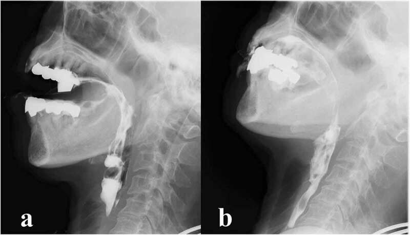
Figure 2.

Videofluoroscopic examination of swallowing (VF) of the patient.
VF of the patient in lateral view at 79 months after the onset of symptoms. Fluoroscopic images (a) before swallowing reflex and (b) and during swallowing. (a) Bolus transport from the oral cavity to the pharynx was poor and initiation of the pharyngeal swallow was delayed. (b) Once the swallowing reflex was triggered, the bolus passed through the penetration pharynx into the upper oesophagus without laryngeal or aspiration
