Figure 1.

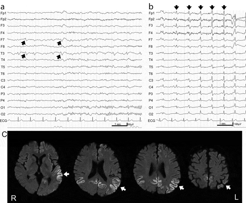
Electroencephalogram (EEG) and magnetic resonance imaging (MRI) in MM2c-sporadic Creutzfeldt-Jakob disease.
(a) EEG readings in the early stage (10 months after disease onset) showed background activity of 10–12 Hz and 75–100 μV and focal discharges (arrows) at F7 and T3, corresponding to hyperintense lesions detected by MRI. (b) EEG readings in the late stage (58 months after disease onset) showed low amplitude background activity at 9–10 Hz and periodic sharp wave complexes at a frequency of 1 Hz (arrows). (c) Diffusion-weighted MRI in the early stage (7 months after disease onset) showed multiple focal hyperintense signals (left-dominant temporo-occipital and bilateral medial occipital cortices in this case; arrows).
