
Figure 2.
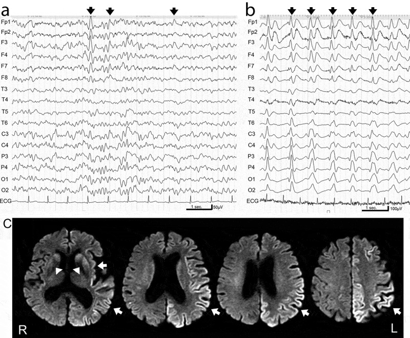
Electroencephalogram (EEG) and magnetic resonance imaging (MRI) in MM1-sporadic Creutzfeldt-Jakob disease.
(a) EEG readings in the early stage (1 month after disease onset) showed background activity of 6–8 Hz and 50–75 μV and lateralized frontal dominant sharp or triphasic waves (arrows). (b) EEG readings in the later stage (4 months after disease onset) showed poorly organized background activity and periodic sharp wave complexes at a frequency of 1 Hz (arrows). (c) Diffusion-weighted MRI in the early stage (2 months after disease onset) showed hyperintense signals in the cortices and basal ganglia (arrows).