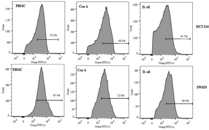
Fig. 9.
Analysis of the mitochondrial membrane depolarization in target cells (HCT-116 and SW620 cells). Target cells were co-cultured with D. oil (10 μg/ml) activated PBMC at 10: 1 (E:T ratio) for 12 h. Mitochondrial damage was analyzed by staining cancer cells with Rh 123 and analyzed by flow cytometry after gating CTFR+ target cells. Untreated PBMC and Con A pretreated PBMC were used as negative and positive controls, respectively. Representative flow diagrams are shown here from two similar experiments

