Figure 4.
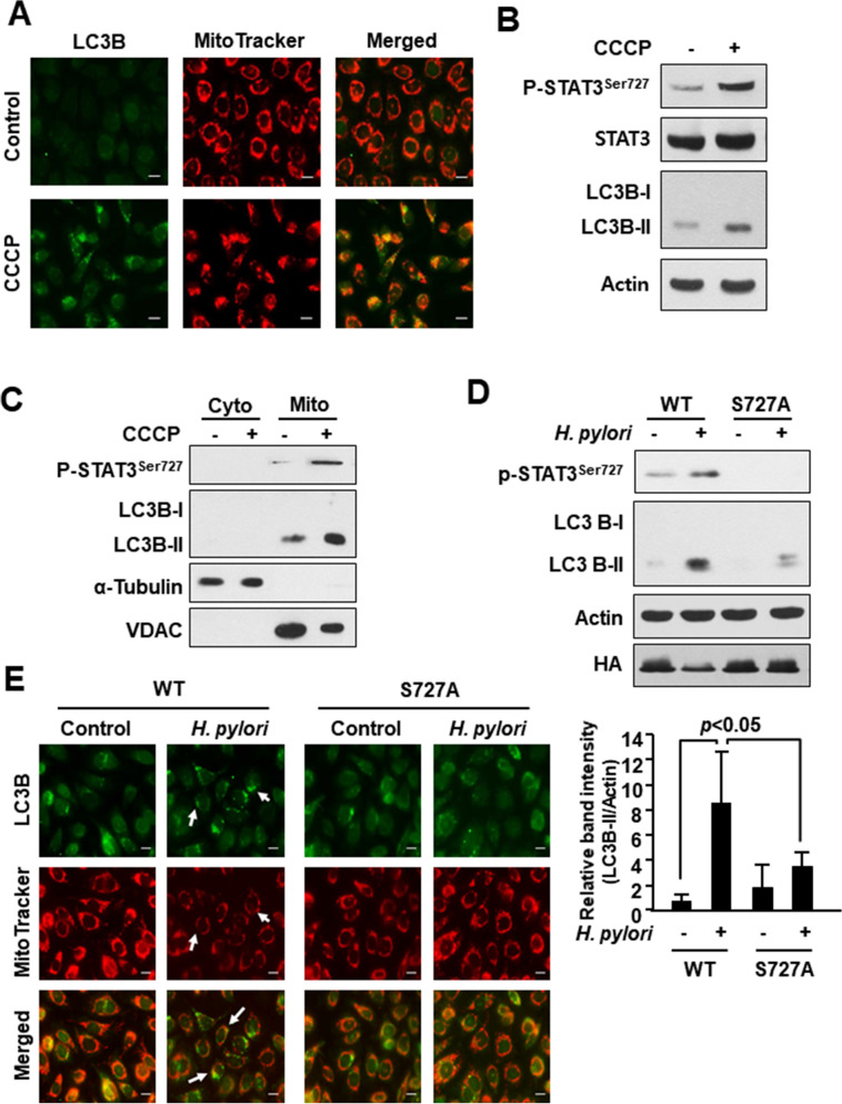
Role of P-STAT3S727 in accumulation of LC3 in mitochondria of H. pylori infected AGS cells. (A) Immunocytochemical analysis of LC3 co-localized with mitochondria. AGS cells were treated with vehicle (DMSO) or CCCP (1 μM) for 1 h. MitoTracker was used as a selective probe of mitochondria. Scale bar, 100 μm. (B) Western blot analysis of P-STAT3Ser727, STAT3, and LC3B in AGS cells treated with DMSO or 1 μM CCCP for 1 h. (C) AGS cells were treated with DMSO or CCCP (1 μM) for 1 h. Western blot analysis of P-STAT3Ser727 and LC3B in cytosolic and mitochondrial extracts. VDAC and α-tubulin are mitochondrial and cytoplasmic protein markers, respectively. (D) Western blot analysis of P-STAT3Ser727 and LC3B in AGS cells transfected with WT or serine dominant negative mutant vector. The relative expression levels of LC3 from three independent experiments are presented means ± S.D. (E) Immunocytochemical analysis of LC3B in WT and Ser727 mutant AGS cells with or without H. pylori infection. MitoTracker is a selective probe of mitochondria. Scale bar, 100 μm.

