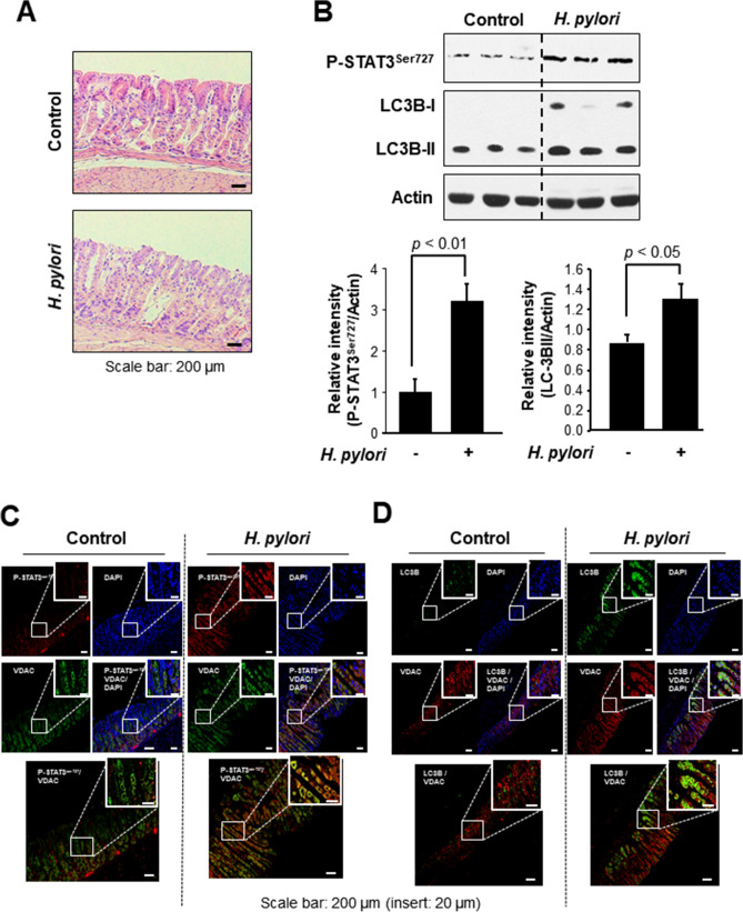
Figure 5.
STAT3 phosphorylation on Ser727 and LC3B expression in mouse stomach infected with H. pylori. (A) H&E staining of H. pylori-infected mouse stomach tissue. (B) Western blot analysis of P-STAT3Ser727 and LC-3B in H. pylori-infected mouse stomach. The relative expression levels of P-STAT3Ser727 and LC-3B are presented means ± S.D. (C) Immunofluorescence analysis of P-STAT3Ser727 co-localized with a mitochondrial marker protein, VDAC. (D) Immunofluorescence analysis of LC3B co-localized with VDAC.

