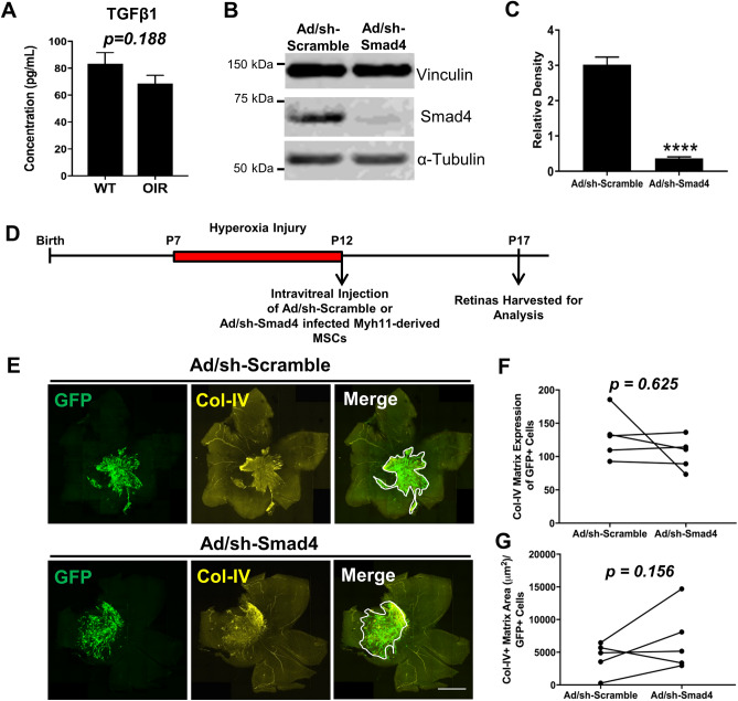
Figure 6.
Smad4 knockdown within Myh11-derived MSCs does not abolish induction of proliferative vitreoretinopathy following intravitreal injection of these cells within the murine OIR model. (A) A Luminex multiplex assay demonstrated no significant difference in active TGFβ1 protein concentration within the neural retina and vitreous of P14 wildtype (WT) mice and WT mice that underwent OIR (n = 5 unpaired eyes). Cropped Western blot from representative lanes of one gel (B) and densitometry quantification (C) demonstrated significant knockdown of Smad4 in Myh11-derived MSCs through mSmad4-shRNA adenovirus vectors (n = 6 biological replicates). Data are represented as mean ± standard error of mean (SEM). (D) Experimental design illustrating the intravitreal injection of Smad4-shRNA infected Myh11-derived MSCs versus Scramble-shRNA infected Myh11-derived MSCs at P12 following OIR injury, with subsequent harvest of injected retinas at P17. (E) Representative tile scan images of immunostained retinas revealed Col-IV pre-retinal matrix production (white outline in Merge panels) in eyes of P17 OIR mice intravitreally injected at P12 with passasge 6–8 Myh11-derived MSCs infected with either Ad-GFP-U6-mSmad4-shRNA or with Ad-GFP-U6-scramble. Fibrotic scar indicated by Col-IV is evident in both eyes regardless of Smad4 knockdown. Scale bar, 1000 µm. (F) No significant difference is found in fibrotic scar Col-IV matrix expression of the eyes intravitreally injected with Myh11-derived MSCs infected with Ad-GFP-U6-scramble adenovirus vectors (GFP+) as compared to Myh11-derived MSCs infected with Ad-GFP-U6-mSmad4-shRNA adenovirus vectors (GFP+) (n = 6 paired eyes). (G) No significant difference is found in fibrotic scar Col-IV matrix area normalized by the number of GFP+ cells found within this fibrotic scar. ****p < 0.0001. Data were analyzed using unpaired t test (A, C), or Wilcoxon test (F, G).

