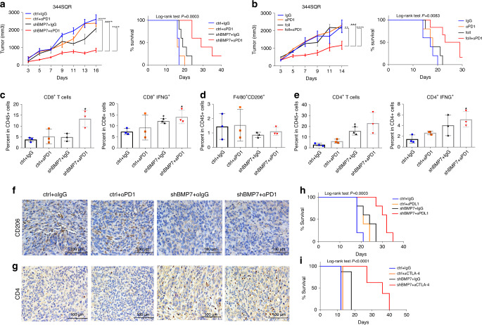
Fig. 5. BMP7 inhibition re-sensitizes resistant tumors to anti-PD1 therapy.
a Tumor growth and survival analysis of mice with 344SQR tumors treated with IgG ctrl (n = 5 animals) or anti-PD1 (10 mg/kg) (n = 5 animals) or 344SQR shBMP7 tumors treated with IgG (n = 5 animals) or anti-PD1 (10 mg/kg) (n = 5 animals) twice a week for 2 weeks. b Tumor growth and survival analysis of mice with 344SQR tumors (n = 5 animals) treated with IgG, anti-PD1 (10 mg/kg), follistatin (0.1 mg/kg), or follistatin (0.1 mg/kg) plus anti-PD1(10 mg/kg) for 2 weeks. For a, ctrl+ IgG vs. shBMP7 + αPD1, ****p < 0.0001, ctrl+ αPD1 vs. shBMP7 + αPD1, ****p < 0.0001 shBMP7+IgG vs. shBMP7 + αPD1, ****p < 0.0001, Two-way RM ANOVA. For b, IgG vs. foll+αPD1, **p = 0.0060, αPD1 vs. foll+αPD1, ***p = 0.0003, foll+IgG vs. foll+αPD1, ****p < 0.0001, Two-way RM ANOVA. Statistical significance was defined as *P < 0.05, **P < 0.01, ***P < 0.001, and ****P < 0.0001. Mouse survival rates were analyzed by the Kaplan–Meier method and compared with log-rank tests. c–e Flow cytometry analysis of CD8+(*p = 0.0421), CD8+IFNG+(*p = 0.0475, *p = 0.0121), F4/80+CD206+, CD4+ (*p = 0.0199), CD4+IFNG+ T cells (*p = 0.0303) in tumor-infiltrating leukocytes (TILs) from 344SQR ctrl (n = 3 biologically independent samples) and 344SQR shBMP7 (n = 3 biologically independent samples) tumors treated with IgG or anti-PD1 (10 mg/kg) twice a week for 2 weeks. Data are presented as mean values ±SD. P values are from unpaired, two-sided t tests. f, g Representative images of immunohistochemical stains for CD206 (M2 macrophage marker) and CD4 (brown dots) in formalin-fixed paraffin-embedded tissue sections from BMP7-knockdown tumors treated with IgG or anti-PD1 compared with control. A representative staining image from each cohort (n = 3 biologically independent samples) is displayed. Data shown are representative of two reproducible independent experiments. Scale bar, 100 μm (×40 magnification). h, i Survival analysis of mice with 344SQR ctrl tumors or 344SQR shBMP7 tumors treated with IgG or anti-PDL1 (10 mg/kg) (n = 5 animals) or anti-CTLA4 (10 mg/kg) (n = 8 animals) twice a week for 2 weeks. Mouse survival rates were analyzed with the Kaplan–Meier method, and curves compared with log-rank tests. Statistical significance was defined as *P < 0.05, **P < 0.01, ***P < 0.001, and ****P < 0.0001.

