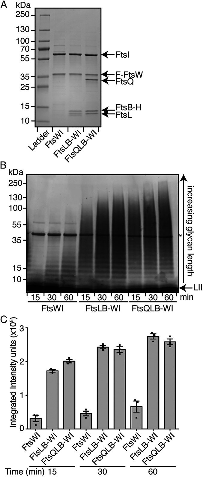
Fig. 1.
FtsQLB stimulates PG polymerization by the FtsWI synthase. (A) Coomassie- stained gel of purified FLAG(F)-tagged FtsW coexpressed with 1) FtsI alone; 2) FtsI, FtsL, and His(H)-tagged FtsB; or 3) FtsI, FtsQ, FtsL, and FtsB-H. Protein concentrations in each preparation were normalized, and equivalent volumes of each were loaded on the gel. (B) PGTase assays using the same normalized preparations shown in A. Purified complexes (0.5 µM) were incubated with purified E. faecalis lipid II (LII) (10 µM) and cephalexin (200 µM) to block cross-linking. The biotin-labeled glycan polymers thus produced were detected on blots with IRDye-labeled streptavidin. The asterisk marks the enzyme used for biotinylating the glycans, which itself becomes biotin-labeled. In both panels, representative images of three independent experiments are shown. (C) The accumulation of glycan fragments from three independent replicates of polymerization time courses was quantified using densitometry. Error bars represent SEM.

