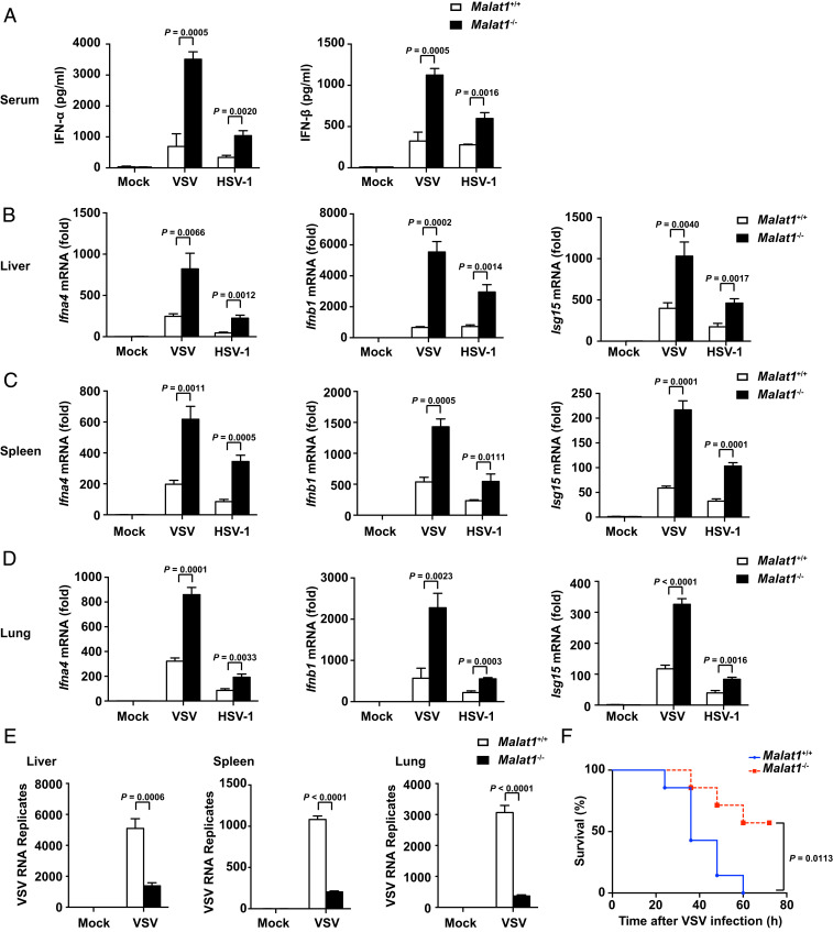
Fig. 3.
Deficiency of Malat1 enhances antiviral response in vivo. (A) ELISA of IFN-α and IFN-β in serum from Malat1+/+ and Malat1−/− mice infected with VSV (5 × 10⁷ pfu/g) for 18 h or HSV-1 (1 × 10⁷ pfu/g) for 24 h, respectively, via tail i.v. injection. (B) qRT-PCR analysis of relative Ifna4, Ifnb1, and Isg15 mRNA level in liver from Malat1+/+ and Malat1−/− mice corresponding to A. (C) qRT-PCR analysis of relative Ifna4, Ifnb1, and Isg15 mRNA level in spleen from Malat1+/+ and Malat1−/− mice corresponding to A. (D) qRT-PCR analysis of relative Ifna4, Ifnb1, and Isg15 mRNA level in lung from Malat1+/+ and Malat1−/− mice corresponding to A. (E) qRT-PCR analysis of relative VSV RNA replication in liver, spleen, and lung from Malat1+/+ and Malat1−/− mice corresponding to A. (F) Survival curves of 6- to 8-wk-old mice of Malat1+/+ and Malat1−/− infected with VSV (1 × 10⁸ pfu/g) via tail i.v. injection. Data are shown as mean ± SD of n = 3 biological replicates (A–E) or demonstrated as Kaplan–Meier survival curve (F, n = 7), two-tailed unpaired Student’s t test (A–E), or Log-rank (Mantel–Cox) test (F).

