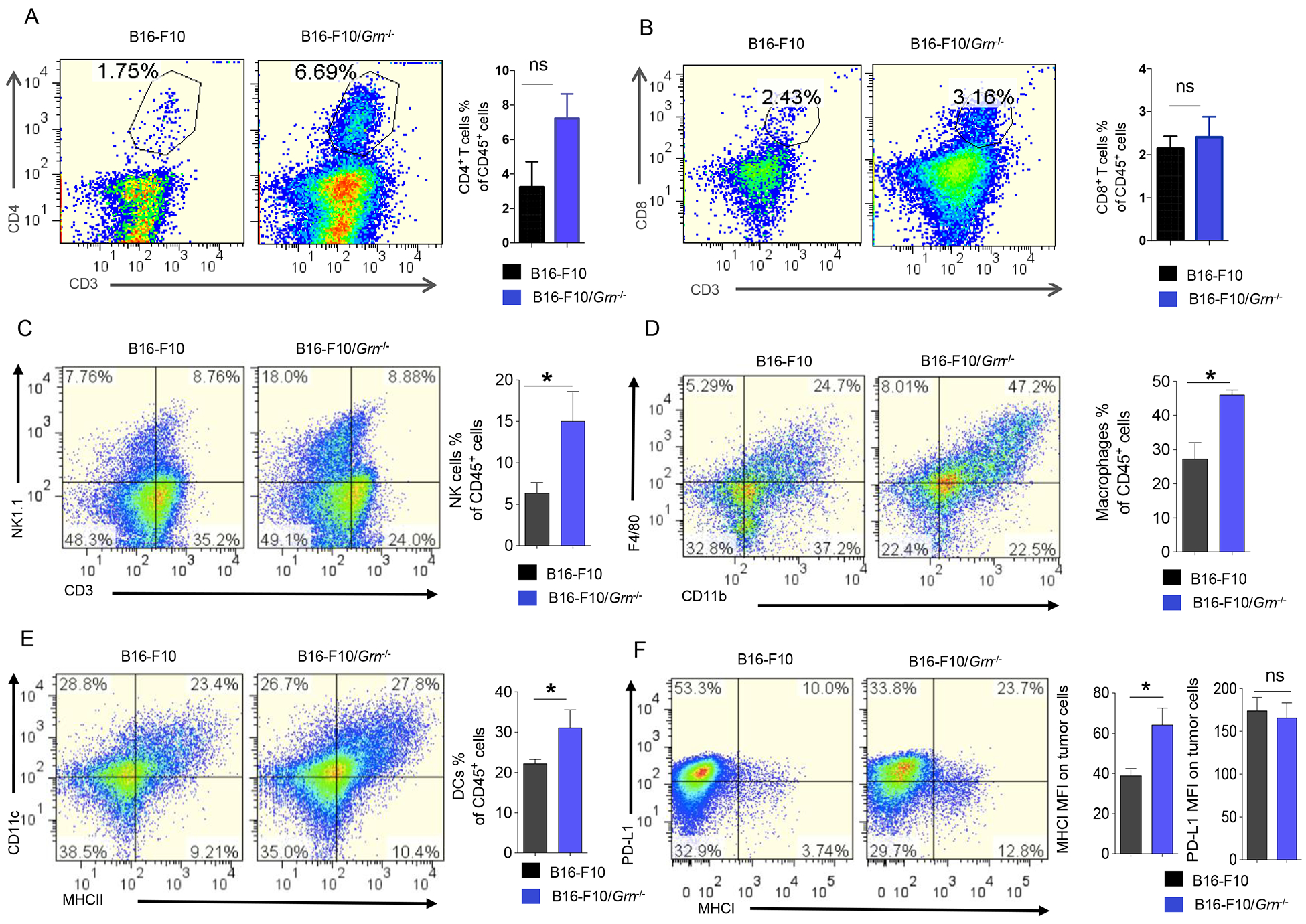
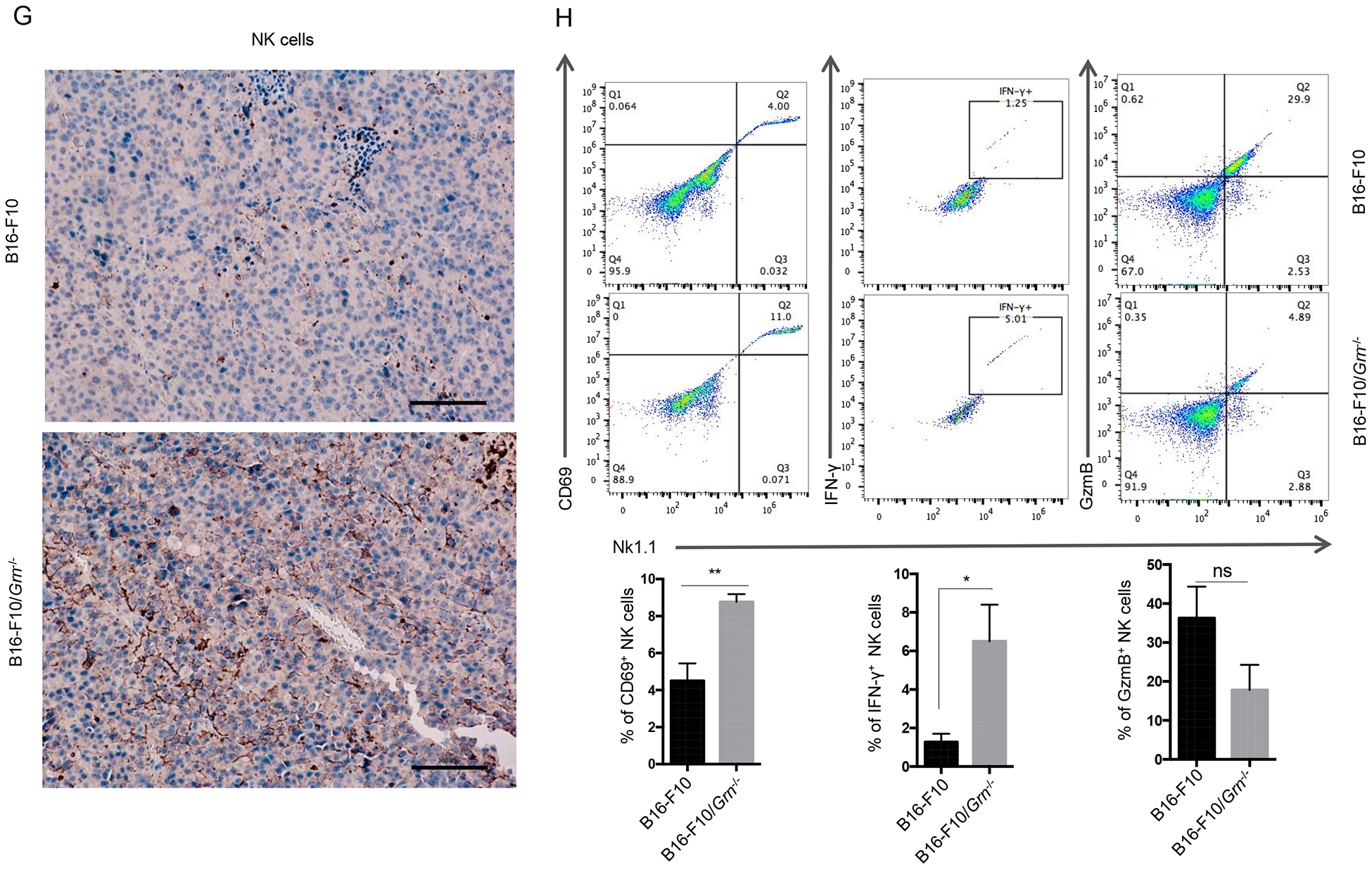
Figure 5. Differential involvement of the immune system in Grn−/− B16-F10 tumor.


1×10⁶ WT, or Grn−/−B16-F10 cells were injected subcutaneously in the right flank to C57BL/6 mice.18 days later, primary tumors were dissected. Single cell suspensions were obtained from these tissues and stained for (A) CD3− NK1.1+ cells, (B) CD11b+ F4/80+ macrophages, (C) CD11c+ MHC II+ dendritic cells, (D) CD4+ T cells, (E) CD8+ T cells. The percentage of each type cells were calculated based on total CD45+ cells. (F) MHC I and PD-L1 expression on the B16-F10/Grn−/− tumors were analyzed by FCM. Data were presented as mean ± SD (* P < 0.05, two-tailed Student’s t test). n=3 mice per group. (G) Immunohistochemical staining for NK cells in ctrl and B16-F10/Grn−/− tumor section by using anti-NK1.1 antibody (Scale bar, 100 μm). (H) Live cells were gated and the gate including CD45+ cells were analyzed for NK1.1, CD69, IFN-γ, and GzmB. Quantification of active NK cells (NK1.1+CD69+, NK1.1+IFN-γ+, NK1.1+GzmB+) infiltrating to the tumor was reported as a percentage of total NK cells. Data were presented as mean ± SEM (* P < 0.05, *** P < 0.001, two-tailed Student’s t test). n=3 mice per group.
