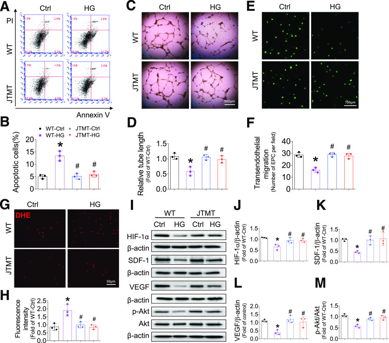
Figure 6.
Endothelial MT overexpression protects EPCs from HG and hypoxia-induced apoptosis and angiogenic dysfunction and impaired HIF-1α/SDF-1/Akt signaling. BM-EPCs from WT and JTMT mice were exposed to HG (25 mmol/L) and hypoxia for 24 h; the equivalent concentration of mannitol was used as osmotic control (Ctrl). The apoptosis was analyzed by flow cytometry using annexin V/PI staining (A and B). The angiogenic function was evaluated by tube formation assay (C and D). The migration capability was evaluated by TEM assay (E and F). The oxidative damage was evaluated by DHE stain of superoxide production (G and H). The expression of HIF-1α (I and J), SDF-1 (I and K), VEGF (I and L), and phosphorylation of Akt (I and M) were tested by Western blot, with β-actin used as loading control. Three independent experiments were performed. Data are mean ± SD. *P < 0.05 vs. WT-Ctrl; #P < 0.05 vs. WT-HG.

