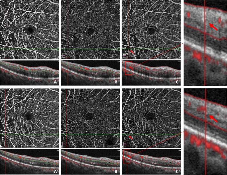
Fig. 4.
Reperfusion occurred in the right eye of a 53-year-old male, 6 months after receiving conbercept treatment. Red arrows and boxes indicate growth of new blood flow signal and reperfusion in the macular nonperfusion areas. OCT angiography images (a–c) at baseline illustrate the macular nonperfusion area, Q-score = 8/10. a Superficial capillary plexus, VD = 45.7%; b Deep capillary plexus, VD = 39.6; c Full retinal capillary plexus. OCT angiography images a’–c’ at month 6 indicate reperfusion in previous macular nonperfusion areas, Q-score = 8/10. a Superficial capillary plexus, VD = 45.5%; b Deep capillary plexus, VD = 40.2%; c Full retinal capillary plexus

