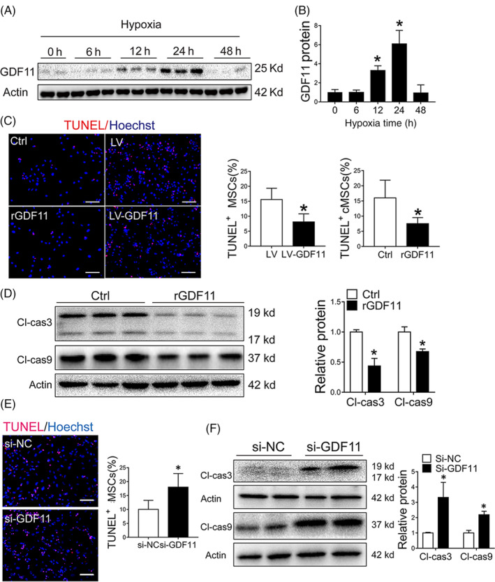
FIGURE 1.

GDF11 protected MSCs against apoptosis under hypoxia condition in vitro. A, GDF11 expression in MSCs were detected by Western blot under normoxia and hypoxia condition at specified times, and β‐actin served as a loading control. B, Quantification of relative GDF11 protein level (n = 3). C, MSCs were pretreated with rGDF11 or overexpressed GDF11 by viral transduction (LV‐GDF11). Representative TUNEL staining images of Control (Ctrl), rGDF11, LV (vector control) and LV‐GDF11 were captured. Scale bar = 50 μm. Quantification of apoptotic cells was presented as ratio of TUNEL‐positive nuclei over the total nuclei from 8 to 10 randomly selected fields in each sample. D, Cleaved caspase 3 (Cl‐cas3) and cleaved caspase 9 (Cl‐cas9) proteins in MSCs pretreated with rGDF11 were assessed by Western blot and quantified by densitometry (n = 3). E, Representative TUNEL staining images of MSCs transfected with siRNA GDF11 (si‐GDF11) or siRNA control (si‐NC). Scale bar = 50 μm. Quantification of apoptotic cells was presented as ratio of TUNEL‐positive nuclei over the total nuclei from 8 to 10 randomly selected fields in each sample. F, Cleaved caspase 3 and 9 proteins of MSCs after transfected with si‐NC and si‐GDF11 were assessed by Western blot; and protein expression levels were quantified by densitometry (n = 4). Data are shown as mean ± SD. *P < .05 vs Ctrl/si‐NC
