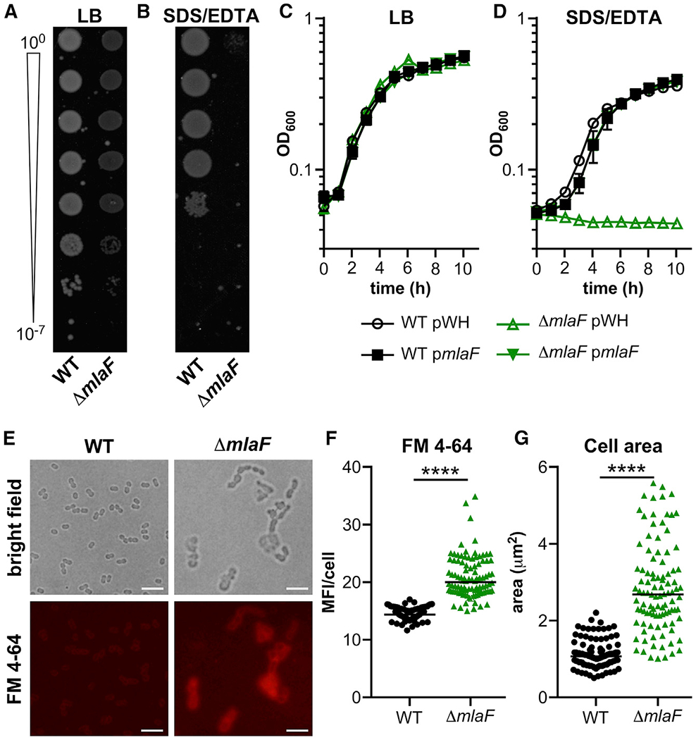
Figure 1. A. baumannii ΔmlaF Has a Growth Defect When Subjected to Membrane Stress and Increased Surface-Accessible Glycero-phospholipids.

(A and B) Wild-type (WT) and ΔmlaF strains were diluted and spotted to lysogeny broth (LB) plates without (A) and with (B) 0.01% SDS and 0.15 mM EDTA.
(C and D) WT and ΔmlaF strains were grown in LB without (C) and with (D) 0.01% SDS and 0.15 mM EDTA (n = 3; data are means ± SEM).
(E) Live WT and ΔmlaF bacteria were incubated with the lipophilic fluorescent dye FM4-64 and imaged on agar pads. Scale bar is 5 μm.
(F and G) The mean fluorescence intensity (MFI) per cell was measured (F).
(G) The cell area (μm2) was measured.
n = 100 with median; significance by Mann-Whitney test. ****p < 0.0001 (F and G). See also Figure S1.
